Korean Circ J.
2011 Jun;41(6):304-307. 10.4070/kcj.2011.41.6.304.
Fractional Flow Reserve Versus Angiography in Left Circumflex Ostial Intervention After Left Main Crossover Stenting
- Affiliations
-
- 1Department of Internal Medicine, Keimyung University College of Medicine, Dongsan Medical Center, Daegu, Korea. shur@dsmc.or.kr
- 2Department of Internal Medicine, Seoul National University College of Medicine, Seoul, Korea.
- 3Department of Internal Medicine, Inje University College of Medicine, Ilsan Paik Hospital, Goyang, Korea.
- 4Stanford University Medical Center, Cardiovascular Medicine, California, USA.
- 5Department of Internal Medicine, Ajou University School of Medicine, Suwon, Korea.
- KMID: 1776186
- DOI: http://doi.org/10.4070/kcj.2011.41.6.304
Abstract
- BACKGROUND AND OBJECTIVES
Discrepancy between angiographic percent (%) diameter stenosis and fractional flow reserve (FFR) exists in non-left main bifurcation lesions. The aim of this study was to compare angiographic stenosis severity and FFR in jailed ostial left circumflex artery (LCX) lesions after left main (LM)-to-left anterior descending artery (LAD) crossover stenting.
SUBJECTS AND METHODS
Twenty-nine (n=29) patients with distal LM or ostial LAD lesions treated by LM-to-LAD crossover stenting were consecutively enrolled. After successful stenting, FFR was measured at the jailed LCX. Additional intervention was performed in lesions with FFR <0.8.
RESULTS
The mean reference diameter of LCX was 3.1+/-0.4 mm, and percent diameter stenosis after crossover stenting was 56+/-21%. Angiographically significant stenosis (>50%) at the ostial LCX occurred in 59% (17/29) of cases. Among them, only five (29%) lesions had functional significance, and underwent additional procedure. During follow-up, three patients in the deferral group and two patients in the additional intervention group had target lesion revascularization.
CONCLUSION
There was a discrepancy between angiographic percent diameter stenosis and FFR in jailed LCX lesions after LM crossover stenting.
Keyword
Figure
-
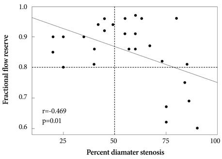
Fig. 1 Correlation between fractional flow reserve and percent diameter stenosis of ostial circumflex coronary artery after left main to left anterior descending coronary artery stenting.
Reference
-
1. Kim YH, Park SW, Hong MK, et al. Comparison of simple and complex stenting techniques in the treatment of unprotected left main coronary artery bifurcation stenosis. Am J Cardiol. 2006. 97:1597–1601.2. Kim W, Kim YJ, Lee WJ, et al. Lesion location: its impacts on the procedural and postprocedural outcomes of unprotected left main coronary stenting. Korean Circ J. 2007. 37:419–424.3. Lim MJ, Kern MJ. Utility of coronary physiologic hemodynamics for bifurcation, aortoostial, and ostial branch stenoses to guide treatment decisions. Catheter Cardiovasc Interv. 2005. 65:461–468.4. Koo BK, Kang HJ, Youn TJ, et al. Physiologic assessment of jailed side branch lesions using fractional flow reserve. J Am Coll Cardiol. 2005. 46:633–637.5. Bellenger NG, Swallow R, Wald DS, et al. Haemodynamic significance of ostial side branch nipping following percutaneous intervention at bifurcations: a pressure wire pilot study. Heart. 2007. 93:249–250.6. King SB 3rd, Smith SC Jr, Hirshfeld JW Jr, et al. 2007 Focused Update of the ACC/AHA/SCAI 2005 Guideline Update for Percutaneous Coronary Intervention: a report of the American College of Cardiology/American Heart Association Task Force on Practice Guidelines: 2007 Writing Group to Review New Evidence and Update the ACC/AHA/SCAI 2005 Guideline Update for Percutaneous Coronary Intervention, Writing on Behalf of the 2005 Writing Committee. Circulation. 2008. 117:261–295.7. Suh JW, Koo BK, Jo SH, et al. Optimal dosage and method of administration of adenosine for measuring the coronary flow reserve and the fractional flow reserve in Koreans. Korean Circ J. 2006. 36:300–307.8. Colombo A, Moses JW, Morice MC, et al. Randomized study to evaluate sirolimus-eluting stents implanted at coronary bifurcation lesions. Circulation. 2004. 109:1244–1249.9. Toyofuku M, Kimura T, Morimoto T, et al. Three-year outcomes after sirolimus-eluting stent implantation for unprotected left main coronary artery disease: insights from the j-Cypher registry. Circulation. 2009. 120:1866–1874.10. White CW, Wright CB, Doty DB, et al. Does visual interpretation of the coronary angiogram predict the physiologic importance of a coronary stenosis. N Engl J Med. 1984. 310:819–824.11. Marcus ML, Skorton DJ, Johnson MR, Collins SM, Harrison DG, Kerter RE. Visual estimate of percent diameter coronary stenosis: a battered gold standard. J Am Coll Cardiol. 1988. 11:882–885.12. Ziaee A, Parham WA, Herrmann SC, Stewart RE, Lim MJ, Kern MJ. Lack of relation between imaging and physiology in ostial coronary artery narrowings. Am J Cardiol. 2004. 93:1404–1407.13. Koo BK, Park KW, Kang HJ, et al. Physiological evaluation of the provisional side-branch intervention strategy for bifurcation lesions using fractional flow reserve. Eur Heart J. 2008. 29:726–732.
- Full Text Links
-
- Actions
-
Cited
- CITED
-
- Close
- Share
-
- Similar articles
-
- Optimal Technique for Ostial Left Anterior Descending Coronary Artery Lesion
- Long-term outcomes of simple crossover stenting from the left main to the left anterior descending coronary artery
- Isolated Coronary Ostial Stenosis Confirmed by Transesophageal Echocardiogram: A Case Report
- A Case of Successful Percutaneous Coronary Intervention by Fractional Flow Reserve and 13N-Ammonia Positron Emission Tomography
- Isolated Ostial left Main Stenosis Diagnosed by Transesophageal Doppler Echocardiography
