J Korean Med Sci.
2014 Apr;29(4):609-613. 10.3346/jkms.2014.29.4.609.
Pneumonectomy Case in a Newborn with Congenital Pulmonary Lymphangiectasia
- Affiliations
-
- 1Department of Pediatrics, Eulji University College of Medicine, Daejeon, Korea. dunggiduk@eulji.ac.kr
- 2Department of Thorasic and Cardiovascular Surgery, Eulji University College of Medicine, Daejeon, Korea.
- 3Department of Pathology, Eulji University College of Medicine, Daejeon, Korea.
- 4Department of Radiology, Eulji University College of Medicine, Daejeon, Korea.
- KMID: 1774471
- DOI: http://doi.org/10.3346/jkms.2014.29.4.609
Abstract
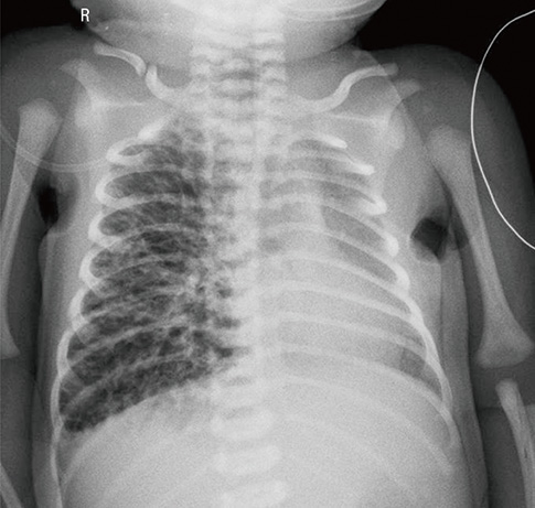
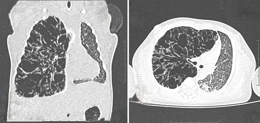
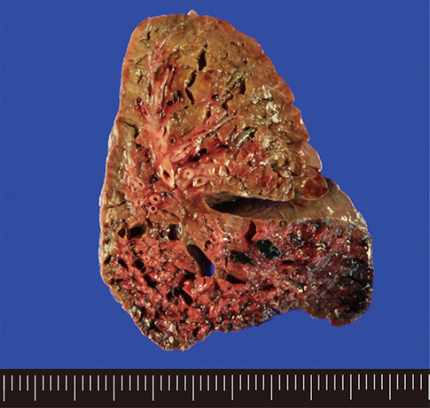
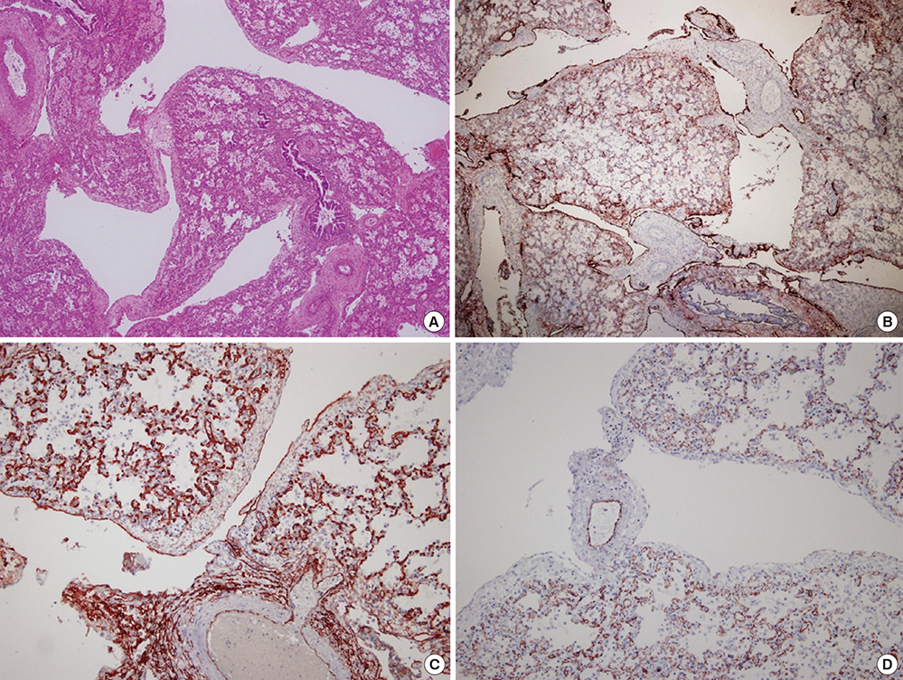
- Congenital pulmonary lymphangiectasia (CPL) is a rare lymphatic pulmonary abnormality. CPL with respiratory distress has a poor prognosis, and is frequently fatal in neonates. We report a case of pneumonectomy for CPL in a newborn. An infant girl, born at 39 weeks' after an uncomplicated pregnancy, exhibited respiratory distress 1 hr after birth, which necessitated intubation and aggressive ventilator care. Right pneumonectomy was performed after her symptoms worsened. Histologic examination indicated CPL. She is currently 12 months old and developing normally. Pneumonectomy can be considered for treating respiratory symptoms for improving chances of survival in cases with unilateral CPL.
MeSH Terms
Figure
Reference
-
1. Esther CR Jr, Barker PM. Pulmonary lymphangiectasia: diagnosis and clinical course. Pediatr Pulmonol. 2004; 38:308–313.2. Virchow R. Gesammelte abdhandlungen zur Wissenschaftliche Medicin. Frankfurt: Meidinger, Sohn & Co.;1856. p. 982.3. Laurence KM. Congenital pulmonary lymphangiectasis. J Clin Pathol. 1959; 12:62–69.4. Mettauer N, Agrawal S, Pierce C, Ashworth M, Petros A. Outcome of children with pulmonary lymphangiectasis. Pediatr Pulmonol. 2009; 44:351–357.5. Bouchard S, Di Lorenzo M, Youssef S, Simard P, Lapierre JG. Pulmonary lymphangiectasia revisited. J Pediatr Surg. 2000; 35:796–800.6. Scott C, Wallis C, Dinwiddie R, Owens C, Coren M. Primary pulmonary lymphangiectasis in a premature infant: resolution following intensive care. Pediatr Pulmonol. 2003; 35:405–406.7. Akcakus M, Koklu E, Bilgin M, Kurtoglu S, Altunay L, Canpolat M, Budak N. Congenital pulmonary lymphangiectasia in a newborn: a response to autologous blood therapy. Neonatology. 2007; 91:256–259.8. Brown M, Pysher T, Coffin CM, Brown M, Pysher T, Coffin CM. Lymphangioma and congenital pulmonary lymphangiectasis: a histologic, immunohistochemical, and clinicopathologic comparison. Mod Pathol. 1999; 12:569–575.9. Rettwitz-Volk W, Schlösser R, Ahrens P, Hörlin A. Congenital unilobar pulmonary lymphangiectasis. Pediatr Pulmonol. 1999; 27:290–292.10. Moore KL. Persaud TVN: the developing human: clinically oriented embryology. 7th ed. Philadelphia: Saunders;2003.11. Noonan JA, Walters LR, Reeves JT. Congenital pulmonary lymphangiectasis. Am J Dis Child. 1970; 120:314–319.12. Laurence KM. Congenital pulmonary cystic lymphangiectasis. J Pathol Bacteriol. 1955; 70:325–333.13. Faul JL, Berry GJ, Colby TV, Ruoss SJ, Walter MB, Rosen GD, Raffin TA. Thoracic lymphangiomas, lymphangiectasis, lymphangiomatosis, and lymphatic dysplasia syndrome. Am J Respir Crit Care Med. 2000; 161:1037–1046.14. Chapdelaine J, Beaunoyer M, St-Vil D, Oligny LL, Garel L, Bütter A, Di Lorenzo M. Unilobar congenital pulmonary lymphangiectasis mimicking congenital lobar emphysema: an underestimated presentation? J Pediatr Surg. 2004; 39:677–680.15. Fukunaga M. Expression of D2-40 in lymphatic endothelium of normal tissues and in vascular tumours. Histopathology. 2005; 46:396–402.16. Jabra AA, Fishman EK, Shehata BM, Perlman EJ. Localized persistent pulmonary interstitial emphysema: CT findings with radiographic-pathologic correlation. AJR Am J Roentgenol. 1997; 169:1381–1384.17. Yamada S, Hisaoka M, Hamada T, Araki S, Shiraishi M. Congenital pulmonary lymphangiectasis: report of an autopsy case masquerading as pulmonary interstitial emphysema. Pathol Res Pract. 2010; 206:522–526.18. Fujishiro J, Komuro H, Ono K, Urita Y, Shinkai T, Minami Y, Kawabata Y, Kishimoto H, Masumoto K. Massive pneumatic expansion of lymphatic vessel resulting in cystic lesions in the pulmonary parenchyma: a rare case of persistent interstitial pulmonary emphysema in a non-ventilated infant. J Pediatr Surg. 2012; 47:e21–e25.19. Hirano H, Nishigami T, Okimura A, Nakasho K, Uematsu K. Autopsy case of congenital pulmonary lymphangiectasis. Pathol Int. 2004; 54:532–536.20. Yalcin S, Ciftci A, Karnak I, Ekinci S, Tanyel FC, Senocak M. Childhood pneumonectomies: two decades' experience of a referral center. Eur J Pediatr Surg. 2013; 23:115–120.
- Full Text Links
- Actions
-
Cited
- CITED
-
- Close
- Share
- Similar articles
-
- A Case of Congenital Pulmonary Lymphangiectasia in Noonan Syndrome
- Congenital Pulmonary Lymphangiectasia, Associated with Total Anomalous Pulmonary Venous Return
- Idiopathic Intestinal Lymphangiectasia
- The Update of Treatment for Primary Intestinal Lymphangiectasia
- Follow up Study of Pneumonectomy for Pulmonary Tuberculosis


