Korean J Gastroenterol.
2012 Dec;60(6):377-381. 10.4166/kjg.2012.60.6.377.
A Case of a Perivascular Epithelioid Cell Tumor Mimicking Colon Cancer
- Affiliations
-
- 1Department of Gastroenterology, Asan Medical Center, University of Ulsan College of Medicine, Seoul, Korea. capsulendos@gmail.com
- KMID: 1773075
- DOI: http://doi.org/10.4166/kjg.2012.60.6.377
Abstract
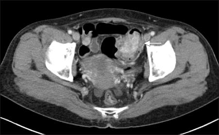
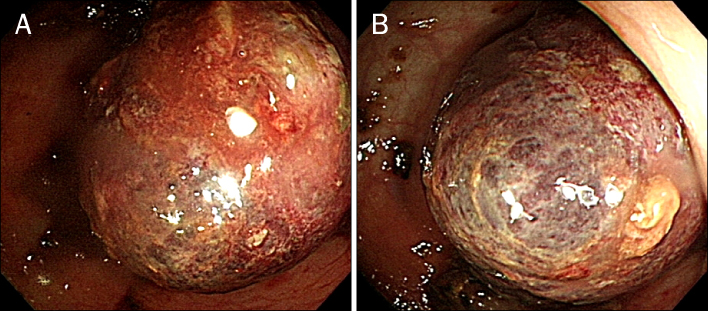
- Perivascular epithelioid cell tumor (PEComa) is extremely rare, which originated from mesenchymal cells in the intestine, and composed of histologically and immunohistochemically distinctive perivascular epithelioid cells. We report here on a case of PEComa in the sigmoid colon. A 62-year-old woman presented with hematochzia 10 days ago. Her abdominal computed tomography scan showed a 5 cm sized intraluminal fungating heterogeneously enhanced, high density mass, which infiltrated pericolic tissue surrounding the sigmoid colon. Colonoscopy showed a purple colored polypoid mass with lobulating contour in the sigmoid colon. She underwent laparoscopic anterior resection. On the histologic examination, the tumor consisted of polygonal epithelioid cells with sheet-like growth of nests, which looked like alveolar tissues in lung. The tumor cells were strongly positive stained with human melanoma black-45 (HMB-45). Pathologic examination was compatible with PEComa. Sixteen months after surgery, she did well without tumor recurrence after surgery. We review the literatures concerning PEComa of the intestine focusing on endoscopic findings.
MeSH Terms
Figure
Reference
-
1. Folpe AL, Kwiatkowski DJ. Perivascular epithelioid cell neoplasms: pathology and pathogenesis. Hum Pathol. 2010. 41:1–15.2. Prasad ML, Keating JP, Teoh HH, et al. Pleomorphic angiomyolipoma of digestive tract: a heretofore unrecognized entity. Int J Surg Pathol. 2000. 8:67–72.3. Kim KH, Jang BI, Kim TN, et al. A case of perivascular epithelioid cell tumor (PEComa) arising in the colon. Korean J Med. 2007. 72:540–545.4. Park SJ, Han DK, Baek HJ, et al. Perivascular epithelioid cell tumor (PEComa) of the ascending colon: the implication of IFN-α 2b treatment. Korean J Pediatr. 2010. 53:975–978.5. Tazelaar HD, Batts KP, Srigley JR. Primary extrapulmonary sugar tumor (PEST): a report of four cases. Mod Pathol. 2001. 14:615–622.6. Bonetti F, Martignoni G, Colato C, et al. Abdominopelvic sarcoma of perivascular epithelioid cells. Report of four cases in young women, one with tuberous sclerosis. Mod Pathol. 2001. 14:563–568.7. Yanai H, Matsuura H, Sonobe H, Shiozaki S, Kawabata K. Perivascular epithelioid cell tumor of the jejunum. Pathol Res Pract. 2003. 199:47–50.8. Birkhaeuser F, Ackermann C, Flueckiger T, et al. First description of a PEComa (perivascular epithelioid cell tumor) of the colon: report of a case and review of the literature. Dis Colon Rectum. 2004. 47:1734–1737.9. Genevay M, Mc Kee T, Zimmer G, Cathomas G, Guillou L. Digestive PEComas a solution when the diagnosis fails to "fit". Ann Diagn Pathol. 2004. 8:367–372.10. Evert M, Wardelmann E, Nestler G, Schulz HU, Roessner A, Röcken C. Abdominopelvic perivascular epithelioid cell sarcoma (malignant PEComa) mimicking gastrointestinal stromal tumour of the rectum. Histopathology. 2005. 46:115–117.11. Yamamoto H, Oda Y, Yao T, et al. Malignant perivascular epithelioid cell tumor of the colon: report of a case with molecular analysis. Pathol Int. 2006. 56:46–50.12. Agaimy A, Wünsch PH. Perivascular epithelioid cell sarcoma (malignant PEComa) of the ileum. Pathol Res Pract. 2006. 202:37–41.13. Baek JH, Chung MG, Jung DH, Oh JH. Perivascular epithelioid cell tumor (PEComa) in the transverse colon of an adolescent: a case report. Tumori. 2007. 93:106–108.14. Pisharody U, Craver RD, Brown RF, Gardner R, Schmidt-Sommerfeld E. Metastatic perivascular epithelioid cell tumor of the colon in a child. J Pediatr Gastroenterol Nutr. 2008. 46:598–601.15. Righi A, Dimosthenous K, Rosai J. PEComa: another member of the MiT tumor family? Int J Surg Pathol. 2008. 16:16–20.16. Tanaka M, Kato K, Gomi K, et al. Perivascular epithelioid cell tumor with SFPQ/PSF-TFE3 gene fusion in a patient with advanced neuroblastoma. Am J Surg Pathol. 2009. 33:1416–1420.17. Qu GM, Hu JC, Cai L, Lang ZQ. Perivascular epithelioid cell tumor of the cecum: a case report and review of literatures. Chin Med J (Engl). 2009. 122:1713–1715.18. Ryan P, Nguyen VH, Gholoum S, et al. Polypoid PEComa in the rectum of a 15-year-old girl: case report and review of PEComa in the gastrointestinal tract. Am J Surg Pathol. 2009. 33:475–482.19. Gross E, Vernea F, Weintraub M, Koplewitz BZ. Perivascular epithelioid cell tumor of the ascending colon mesentery in a child: case report and review of the literature. J Pediatr Surg. 2010. 45:830–833.20. Folpe AL, Mentzel T, Lehr HA, Fisher C, Balzer BL, Weiss SW. Perivascular epithelioid cell neoplasms of soft tissue and gynecologic origin: a clinicopathologic study of 26 cases and review of the literature. Am J Surg Pathol. 2005. 29:1558–1575.
- Full Text Links
- Actions
-
Cited
- CITED
-
- Close
- Share
- Similar articles
-
- Perivascular epithelioid cell tumor (PEComa) of the ascending colon: the implication of IFN-alpha2b treatment
- Malignant Perivascular Epithelioid Cell Tumor (PEComa) Arising in the Omentum with Metastatic Peritoneal Seeding: A Case Report
- Primary Perivascular Epithelioid Cell Tumor (PEComa) of the Liver: A Case Report and Review of the Literature
- A Case of Primary Cutaneous Perivascular Epithelioid Cell Tumor
- A case of perivascular epithelioid cell tumor (PEComa) arising in the colon


