Air Bubbles Mimic Disc Herniation in MRI after Cervical Epidural Block
- Affiliations
-
- 1Department of Anesthesiology and Pain Medicine, Sae-Woori Spine Hospital, Gwangju, Korea. drfirst@hanmail.net
- 2Department of Anesthesiology and Pain Medicine, Christian Hospital, Gwangju, Korea.
- KMID: 1767883
- DOI: http://doi.org/10.3344/kjp.2010.23.3.202
Abstract
- Magnetic resonance image (MRI) is the most sensitive imaging test of the spine in routine clinical practice. Unlike conventional x-ray examinations and computed tomography scans, high-quality magnetic resonance images can be assured only if patients are able to remain perfectly still. However, some patients find it uncomfortable to remain still because of pain. In that condition, interlaminar cervical epidural injections can reduce pain and allow the procedure. When using air with the "loss of resistance" technique in epidural injections to identify the epidural space, there is the possibility of injected excessive air epidurally to mimic a herniated disc. We describe a case report of epidural air artifact in a cervical MRI after cervical epidural injections.
MeSH Terms
Figure
-
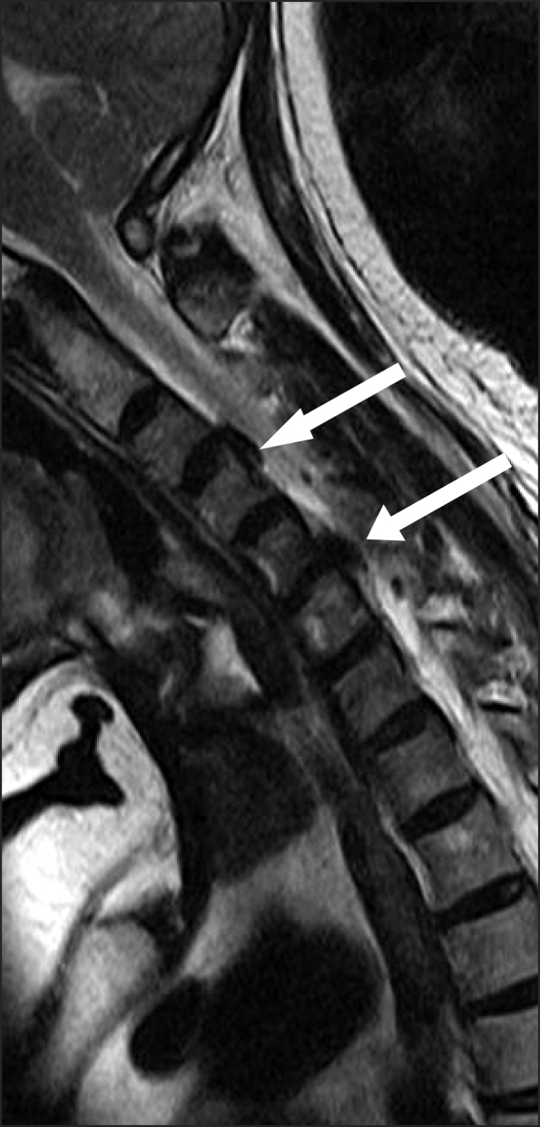
Fig. 1 T2 sagittal image after cervical epidural block shows low signal intensity lesion at C3-C4 and C5-C6. These lesion are considered herniated disc.
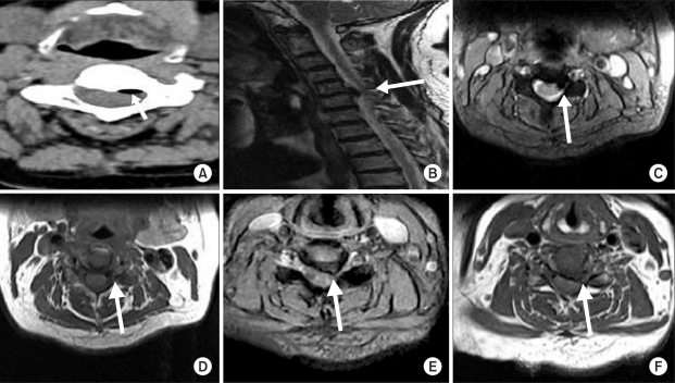
Fig. 2 CT axial image (A) shows low density lesion behind C3-C4 disc. At CT, it seems like the air, not herniated disc. Low signal intensity lesion behind C3-C4 disc seems new compared MRIs before and after cervical epidural block. There is no lesion behind C3-C4 disc in MRI before cervical epidural block (B). (C) and (D) show air as a low signal intensity. By contrast, (E) and (F) show C5-C6 herniated disc relatively higher intensity than lesion behind C3-C4.
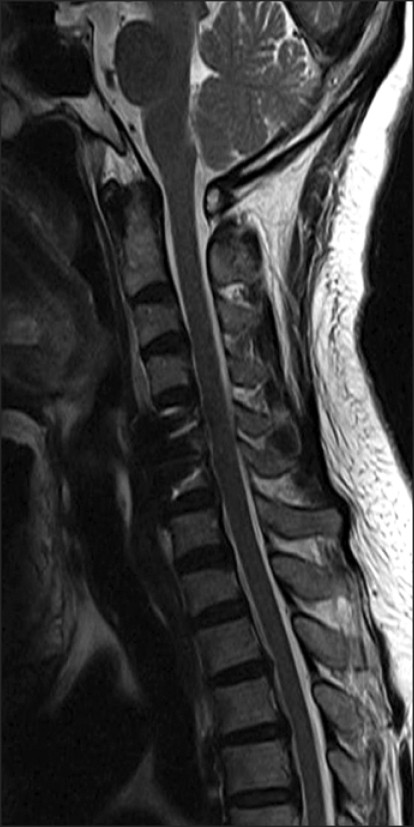
Fig. 3 Post-op. T2 sagittal image. There is no air behind C3-C4 disc.
Cited by 4 articles
-
An Alternative Approach to Needle Placement in Cervicothoracic Epidural Injections
Seung Yong Park, Jung Gil Leem, Sung Hwan Jung, Young Ki Kim, Won Uk Koh
Korean J Pain. 2012;25(3):183-187. doi: 10.3344/kjp.2012.25.3.183.Motor Weakness after Caudal Epidural Injection Using the Air-acceptance Test
Mi Hyeon Lee, Cheol Sig Han, Sang Hoon Lee, Jeong Hyun Lee, Eun Mi Choi, Young Ryong Choi, Mi Hwa Chung
Korean J Pain. 2013;26(3):286-290. doi: 10.3344/kjp.2013.26.3.286.Review of Medical Dispute Cases in the Pain Management in Korea: A Medical Malpractice Liability Insurance Database Study
Yeon Dong Kim, Hyun Seog Moon
Korean J Pain. 2015;28(4):254-264. doi: 10.3344/kjp.2015.28.4.254.Effect of epidural corticosteroid injection on magnetic resonance imaging findings
Min Soo Kim, Tae Yoon Jeong, Yu Seon Cheong, Young Wook Jeon, So Young Lim, Seong Sik Kang, In Nam Kim, Tsong Bin Chang, Hyun Ho Seong, Byeong Mun Hwang
Korean J Pain. 2017;30(4):281-286. doi: 10.3344/kjp.2017.30.4.281.
Reference
-
1. Saberski LR, Kondamuri S, Osinubi OY. Identification of the epidural space: is loss of resistance to air a safe technique? A review of the complications related to the use of air. Reg Anesth. 1997; 22:3–15. PMID: 9010941.
Article2. Mateo E, López-Alarcón MD, Moliner S, Calabuig E, Vivó M, De Andrés J, et al. Epidural and subarachnoidal pneumocephalus after epidural technique. Eur J Anaesthesiol. 1999; 16:413–417. PMID: 10434173.
Article3. Dalens B, Bazin JE, Haberer JP. Epidural bubbles as a cause of incomplete analgesia during epidural anesthesia. Anesth Analg. 1987; 66:679–683. PMID: 3605680.
Article4. Boezaart AP, Levendig BJ. Epidural air-filled bubbles and unblocked segments. Can J Anaesth. 1989; 36:603–604. PMID: 2791185.
Article5. Abbott MA, Samuel JR, Webb DR. Anesthesia for extracorporeal shock wave lithotripsy. Anaesthesia. 1985; 40:1065–1072. PMID: 4073422.
Article6. Kennedy TM, Ullman DA, Harte FA, Saberski LR, Greenhouse BB. Lumbar root compression secondary to epidural air. Anesth Analg. 1988; 67:1184–1186. PMID: 3195736.
Article7. Hirsch M, Katz Y, Sasson A. Spinal cord compression by unusual epidural air accumulation after continuous epidural analgesia. AJR Am J Roentgenol. 1989; 153:887–888. PMID: 2773752.
Article8. Miguel R, Morse S, Murtagh R. Epidural air associated with multiradicular syndrome. Anesth Analg. 1991; 73:92–94. PMID: 1858997.
Article9. Petty R, Stevens R, Erickson S, Lucio J, Kao TC. Inhalation of nitrous oxide expands epidural air bubbles. Reg Anesth. 1996; 21:144–148. PMID: 8829407.10. Stevens R, Mikat-Stevens M, Van Clief M, Schubert A, Weinstein Z. Deliberate epidural air injection in dogs: a radiographic study. Reg Anesth. 1989; 14:180–182. PMID: 2491280.11. Cuerden C, Buley R, Downing JW. Delayed recovery after epidural block in labour. A report of four cases. Anaesthesia. 1977; 32:773–776. PMID: 920919.
- Full Text Links
-
- Actions
-
Cited
- CITED
-
- Close
- Share
-
- Similar articles
-
- Cervicogenic Headache Caused by Cervical C3-C4 Intervertebral Disc Herniation : A case report
- Pneumocephalus after Interlaminar Lumbar Epidural Block
- Unintended subdural anesthesia and subdural air bubbles after attempted epidural anesthesia in a patient undergoing cesarean section
- Posterior Epidural Herniation of a Lumbar Disk Fragment at L2–3 That Mimicked an Epidural Hematoma
- A Case of Cervical Far Lateral Disc Herniation-Case Report-
