Treatment of skeletal Class II adult patient with vertical and transverse problems caused by nasal airway obstruction using microimplant anchorage
- Affiliations
-
- 1Department of Orthodontics, School of Dentistry, Wonkwang University, Korea. jongmoon@wonkwang.ac.kr
- 2The Korean Orthodontic Research Institute Inc., Korea.
- KMID: 1762581
- DOI: http://doi.org/10.4041/kjod.2009.39.4.257
Abstract
- This case report describes the treatment of an adult patient with a Class I canine and molar relationship but a convex profile with a retrognathic mandible and marked lip protrusion, as well as an excessive lower anterior facial height and reduced transverse width on both arches due to a nasal airway obstruction. The constricted arches were expanded by surgically-assisted rapid palatal expansion and the application of a Schwarz appliance to the maxilla and mandible. Acceptable facial balance was obtained using contemporary directional force technology with microimplant anchorage (MIA), which provided horizontal and vertical anchorage in the maxillary and mandibular posterior teeth, as well as intrusion and torque control in the maxillary anterior teeth, resulting in a favorable counterclockwise mandibular response. The total treatment period was 29 months and the results were acceptable for 13 months after debonding.
Keyword
Figure
-
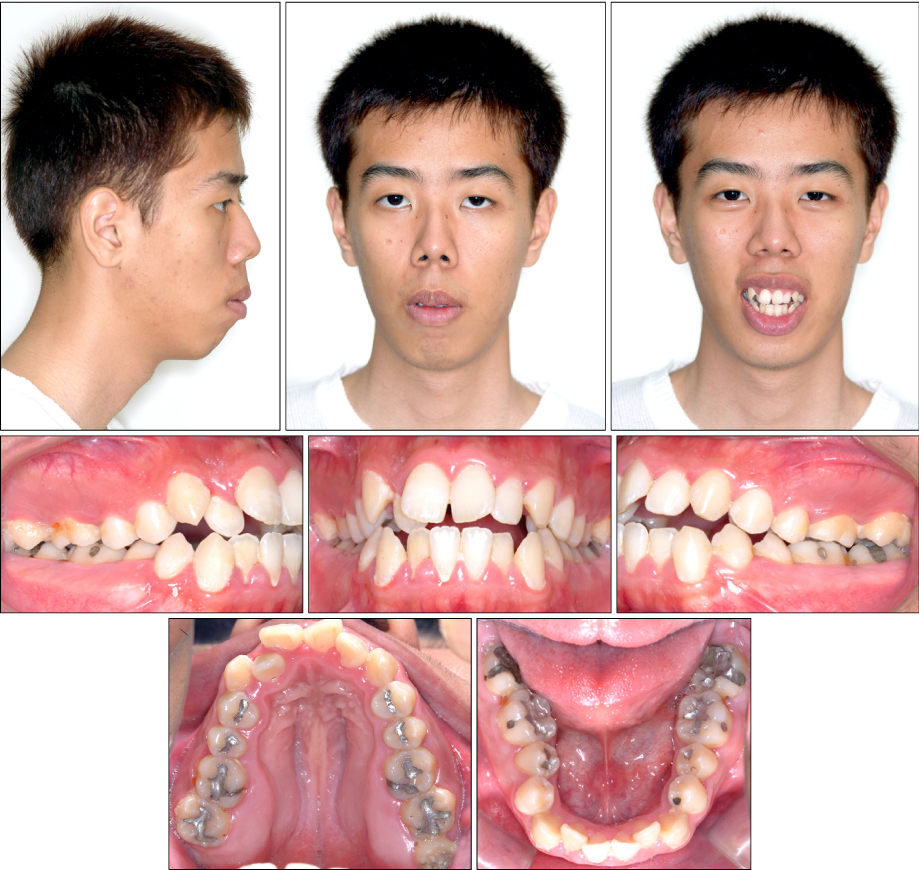
Fig 1 Pretreatment facial and intraoral photographs.

Fig 2 Pretreatment radiographs.

Fig 3 Surgical assisted rapid maxillary expansion on the maxilla and Schwarz appliance on the mandible.
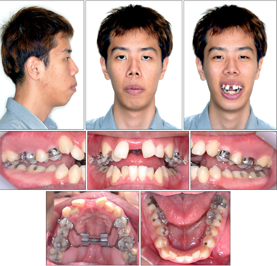
Fig 4 Photographs after 28 day expansion of the maxilla and mandible.
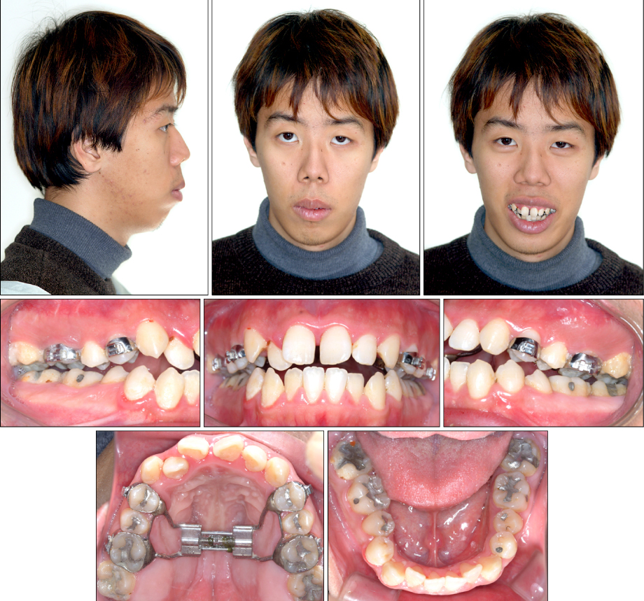
Fig 5 Photographs after 3.5 months of retention of the maxilla and 5 months of expansion of the mandible.
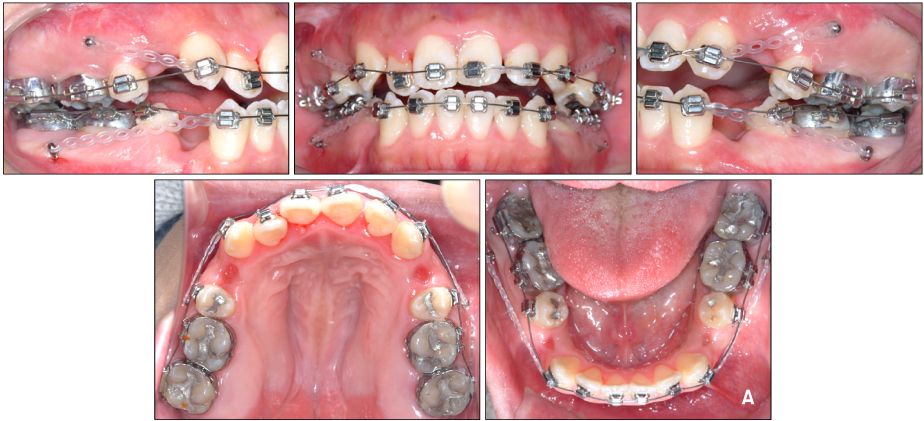
Fig 6. A Denture preparation: retraction of the maxillary and mandibular canines to level six anterior teeth.

Fig 6. B Denture correction: en masse retraction with a closing loop archwire supported by mandibular posterior MIs, intruding forces with elastomeric chain between the main archwires and maxillary and mandibular posterior MIs, and unilateral Class III elastics to correct asymmetry.
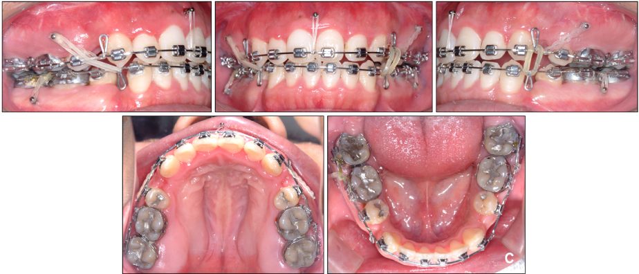
Fig 6. C Denture correction: en masse retraction with closing loop archwire supported by maxillary posterior and anterior microimplants.
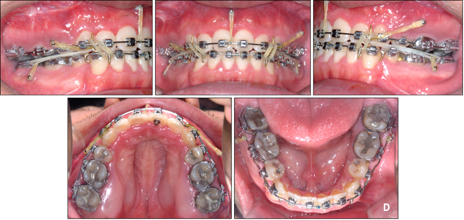
Fig 6. D Denture completion: directional force and finishing with Class II and cusp seating elastics, and elastomeric chain and microimplants.

Fig 7 Post-treatment facial and intraoral photographs.
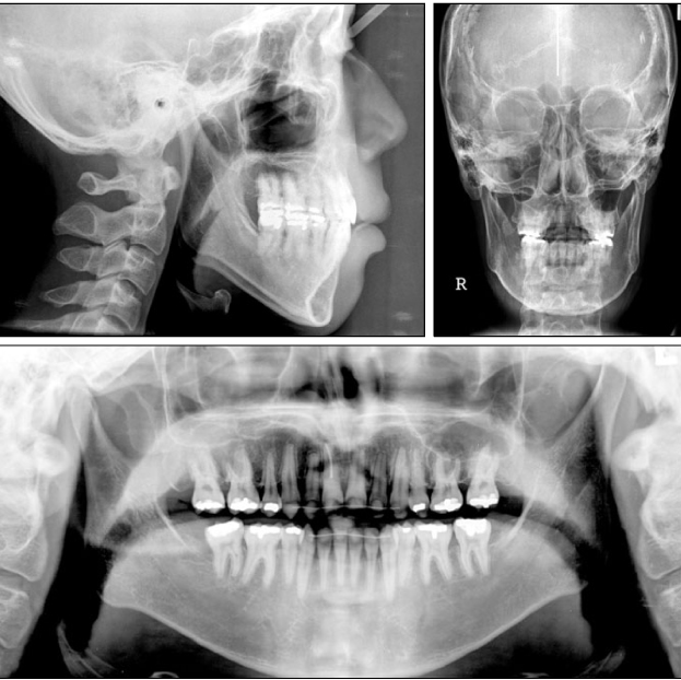
Fig 8 Post-treatment radiographs.
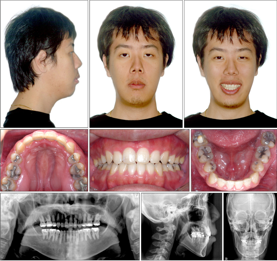
Fig 9 13-month retention photographs and radiographs.
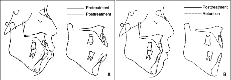
Fig 10 A, Pretreatment and post-treatment cephalometric superimposition; B, post-treatment and 13-month retention cephalometric superimposition.
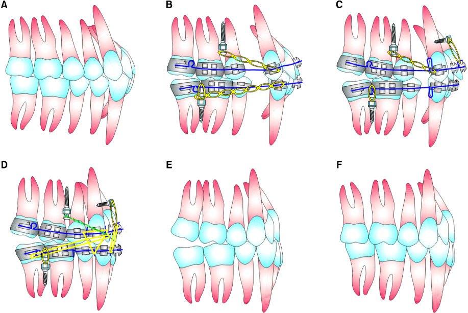
Fig 11 Schematic illustration of the contemporary directional technology using MIA in the case of Class II div.1 bialveolar protrusion. A, Before treatment; B, placement of posterior MIs, canine retraction for leveling in both arches; C, placement of maxillary anterior MI, en masse retraction of six anterior teeth, using loop mechanics in both arches; D, directional forces; E, Tweed occlusion; F, denture recovery.
Cited by 2 articles
-
Non-surgical treatment and retention of open bite in adult patients with orthodontic mini-implants
Cheol-Hyun Moon, Joo-Sin Lee, Hyun-Sun Lee, Jin-Hugh Choi
Korean J Orthod. 2009;39(6):402-419. doi: 10.4041/kjod.2009.39.6.402.Three-dimensional finite element analysis for determining the stress distribution after loading the bone surface with two-component mini-implants of varying length
Bohm Choi, Dong-Ok Lee, Sung-Seo Mo, Seong-Hun Kim, Ki-Ho Park, Kyu-Rhim Chung, Gerald Nelson, Seong Ho Han
Korean J Orthod. 2011;41(6):423-430. doi: 10.4041/kjod.2011.41.6.423.
Reference
-
1. Quinn GW. Airway interference syndrome. Clinical identification and evaluation of nose breathing capabilities. Angle Orthod. 1983. 53:311–319.2. Bresolin D, Shapiro PA, Shapiro GG, Chapko MK, Dassel S. Mouth breathing in allergic children: its relationship to dentofacial development. Am J Orthod. 1983. 83:334–340.
Article3. Schlenker WL, Jennings BD, Jeiroudi MT, Caruso JM. The effects of chronic absence of active nasal respiration on the growth of the skull: a pilot study. Am J Orthod Dentofacial Orthop. 2000. 117:706–713.
Article4. McNamara JA. Influence of respiratory pattern on craniofacial growth. Angle Orthod. 1981. 51:269–300.
Article5. Ricketts RM. Respiratory obstruction syndrome. Am J Orthod. 1968. 54:495–507.6. Northway WM, Meade JB Jr. Surgically assisted rapid maxillary expansion: a comparison of technique, response, and stability. Angle Orthod. 1997. 67:309–320.7. Hartgerink DV, Vig PS, Abbott DW. The effect of rapid maxillary expansion on nasal airway resistance. Am J Orthod Dentofacial Orthop. 1987. 92:381–389.
Article8. Basciftci FA, Mutlu N, Karaman AI, Malkoc S, Küçükkolbasi H. Does the timing and method of rapid maxillary expansion have an effect on the changes in nasal dimensions? Angle Orthod. 2002. 72:118–123.9. Bicakci AA, Agar U, Sökücü O, Babacan H, Doruk C. Nasal airway changes due to rapid maxillary expansion timing. Angle Orthod. 2005. 75:1–6.10. Betts NJ, Vanarsdall RL, Barber HD, Higgins-Barber K, Fonseca RJ. Diagnosis and treatment of transverse maxillary deficiency. Int J Adult Orthodon Orthognath Surg. 1995. 10:75–96.11. Lanigan DT, Mintz SM. Complications of surgically assisted rapid palatal expansion: review of the literature and report of a case. J Oral Maxillofac Surg. 2002. 60:104–110.
Article12. Haas AJ. Rapid expansion of the maxillary dental arch and nasal cavity by opening the mid-palatal suture. Angle Orthod. 1961. 31:73–90.13. Wertz RA. Skeletal and dental changes accompanying rapid midpalatal suture opening. Am J Orthod. 1970. 58:41–66.
Article14. Bishara SE, Staley RN. Maxillary expansion: clinical implications. Am J Orthod Dentofacial Orthop. 1987. 91:3–14.
Article15. Wendling LK, McNamara JA Jr, Franchi L, Baccttti T. A prospective study of the short term treatment effects of the acrylic-splint rapid maxillary expander combined with the lower Schwarz appliance. Angle Orthod. 2004. 75:7–14.16. Vig Ps, Sarver DM, Hall DJ, Warren DW. Quantitative evaluation of nasal airway in relation to facial morphology. Am J Orthod. 1981. 79:263–272.17. Chae JM. A new protocol of Tweed-Merrifield directional force technology with microimplant anchorage. Am J Orthod Dentofacial Orthop. 2006. 130:100–109.
Article18. Park HS, Kwon TG, Kwon OW. Treatment of open bite with microscrew implant anchorage. Am J Orthod Dentofacial Orthop. 2004. 126:627–636.
Article19. Trask GM, Shapiro GG, Shapiro PA. The effects of perennial allergic rhinitis on dental and skeletal development: a comparison of sibling pairs. Am J Orthod Dentofacial Orthop. 1987. 92:286–293.
Article20. Tourne LP. The long face syndrome and impairment of the nasopharyngeal airway. Angle Orthod. 1990. 60:167–176.
Article21. Langford SR, Sims MR. Root surface resorption, repair, and periodontal attachment following rapid maxillary expansion in man. Am J Orthod. 1982. 81:108–115.
Article22. Bays RA, Greco JM. Surgically assisted rapid palatal expansion: an outpatient technique with long-term stability. J Oral Maxillofac Surg. 1992. 50:110–113.
Article23. Berger JL, Pangrazio-Kulbersh V, Borgula T, Kaczynski R. Stability of orthopedic and surgically assisted rapid palatal expansion over time. Am J Orthod Dentofacial Orthop. 1998. 114:638–645.
Article24. Ozturk M, Doruk C, Ozec I, Polat S, Babacan H, Bicakci AA. Pulpal blood flow: effects of corticotomy and midline osteotomy in surgically assisted rapid palatal expansion. J Craniomaxillofac Surg. 2003. 31:97–100.
Article25. Cureton SL, Cuenin M. Surgically assisted rapid palatal expansion: orthodontic preparation for clinical success. Am J Orthod Dentofacial Orthop. 1999. 116:46–59.
Article26. Motoyoshi M, Shirai S, Yano S, Nakanishi K, Shimizu N. Permissible limit for mandibular expansion. Eur J Orthod. 2005. 27:115–120.
Article27. O'Grady PW, McNamara JA Jr, Baccetti T, Franchi L. A long-term evaluation of the mandibular Schwarz appliance and the acrylic splint expander in early mixed dentition patients. Am J Orthod Dentofacial Orthop. 2006. 130:202–213.28. Merrifield LL, Cross JJ. Directional forces. Am J Orthod. 1970. 57:435–464.
Article29. Vanden Bulcke MM, Burstone CJ, Sachdeva RC, Dermaut LR. Location of the center of resistance for anterior teeth during retraction using the laser reflection technique. Am J Orthod Dentofac Orthop. 1987. 91:375–384.
Article30. Jeong GM, Sung SJ, Lee KJ, Chun YS, Mo SS. Finite-element investigation of the center of resistance of the maxillary dentition. Korean J Orthod. 2009. 39:83–94.
Article31. Lee HA, Park YC. Treatment and posttreatment changes following intrusion of maxillary posterior teeth with miniscrew implants for open bite correction. Korean J Orthod. 2008. 38:31–40.
Article32. Sugawara J, Baik UB, Umemori M, Takahashi I, Nagasaka H, Kawamura H, et al. Treatment and posttreatment dentoalveolar changes following intrusion of mandibular molars with application of a skeletal anchorage system (SAS) for open bite correction. Int J Adult Orthod Orthognath Surg. 2002. 17:243–253.33. Reitan K. Principles of retention and avoidance of posttreatment relapse. Am J Orthod. 1969. 55:776–790.
Article34. Reitan K, Kvam E. Comparative behavior of human and animal tissue during experimental tooth movement. Angle Orthod. 1971. 41:1–14.35. Denison TF, Kokich VG, Shapiro PA. Stability of maxillary surgery in open bite versus nonopen bite malocclusions. Angle Orthod. 1989. 59:5–10.36. Proffit WR, Bailey LJ, Phillips C, Turvey TA. Long-term stability of surgical open-bite correction by Le Fort I osteotomy. Angle Orthod. 2000. 70:112–117.
- Full Text Links
-
- Actions
-
Cited
- CITED
-
- Close
- Share
-
- Similar articles
-
- Nonextraction treatment of Class II division 2 in an adult patient using microimplant anchorage (MIA)
- Directional forces using skeletal anchorage for treatment of skeletal Class II div.1 malocclusion
- Microimplant mandibular advancement (MiMA) therapy for the treatment of snoring and obstructive sleep apnea (OSA)
- A new protocol of the sliding mechanics with micro-implant anchorage(M.I.A.)
- Class III nonsurgical treatment using indirect skeletal anchorage: A case report
