Solitary Fibrous Tumor of the Trachea: CT Findings with a Pathological Correlation
- Affiliations
-
- 1Department of Radiology, Gachon University Gil Medical Center, Incheon, Korea. drchoi126@gilhospital.com
- 2Department of Cardiovascular and Thoracic Surgery, Gachon University Gil Medical Center, Incheon, Korea.
- KMID: 1758465
- DOI: http://doi.org/10.3348/kjr.2008.9.3.286
Abstract
- We present the multidetector CT findings with a pathologic correlation for the case of a solitary fibrous tumor located in the trachea. The MDCT revealed a well-circumscribed intraluminal mass arising from the trachea, with strong nodular enhancement in the periphery of the mass. The enhancement pattern of the mass corresponded histopathologically to a focal hypocellular area in the center and prominent blood vessels along the periphery of the mass. We also present volume-rendered and virtual bronchoscopic images of this rare submucosal tracheal tumor.
Keyword
MeSH Terms
Figure
-
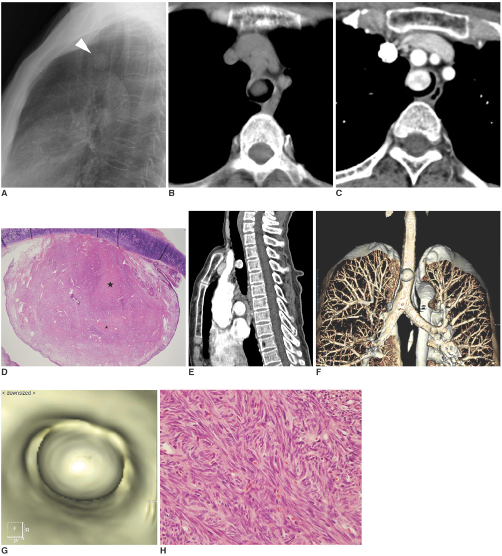
Fig. 1 62-year-old woman with solitary fibrous tumor of trachea. A. Lateral chest radiograph revealing well demarcated mass, with round opacity (arrowhead) in mid level of trachea. B. Non-contrast enhanced CT scan revealing well-defined, round, intraluminal mass with homogeneous soft tissue attenuation in trachea. C. Contrast enhanced CT scan depicting strong enhancement in peripheral portion of mass with central area of low attenuation. D. Photomicrograph (Hematoxylin & Eosin staining, × 10) demonstrating round exophytic submucosal mass with various sized blood vessels in its periphery. It is primarily composed of tumor rich areas with some areas of collagen (asterisks). E. Oblique sagittal image of mass located in mid level of trachea, arising from anterior wall. F, G. Volume-rendered and virtual bronchoscopic images revealing round intraluminal mass arising from anterior tracheal wall. H. Histological examination (Hematoxylin & Eosin staining, × 400) revealed haphazard growth pattern of short spindle cells with scant cytoplasm and strands of rope-like collagen. Immunohistochemical study showed positive response for CD34 and negative response for smooth muscle actin, desmin and S100 protein (not shown), which is consistent with solitary fibrous tumor.
Reference
-
1. Miller WT Jr. Obstructive diseases of the trachea. Semin Roentgenol. 2001. 36:21–40.2. Shah H, Garbe L, Nussbaum E, Dumon JF, Chiodera PL, Cavaliere S. Benign tumors of the tracheobronchial tree. Endoscopic characteristics and role of laser resection. Chest. 1995. 107:1744–1751.3. Ko JM, Jung JI, Park SH, Lee KY, Chung MH, Ahn MI, et al. Benign tumors of the tracheobronchial tree: CT-pathologic correlation. AJR Am J Roentgenol. 2006. 186:1304–1313.4. Gold JS, Antonescu CR, Hajdu C, Ferrone CR, Hussain M, Lewis JJ, et al. Clinicopathologic correlation of solitary fibrous tumors. Cancer. 2002. 94:1057–1068.5. Klemperer P, Rabin CB. Primary neoplasms of the pleura: a report of five cases. Arch Pathol. 1931. 11:385–412.6. England DM, Hochholzer L, McCarthy MJ. Localized benign and malignant fibrous tumors of the pleura: a clinicopathologic review of 223 cases. Am J Surg Pathol. 1989. 13:640–658.7. Moran CA, Suster S, Koss MN. The spectrum of histologic growth patterns in benign and malignant fibrous tumors of the pleura. Semin Diagn Pathol. 1992. 9:169–180.8. Rosade-De-Christenson ML, Abbott GF, McAdams HP, Franks TJ, Galvin JR. From the archives of the AFIP: localized fibrous tumor of the pleura. Radiographics. 2003. 23:759–783.9. Ganly I, Patel SG, Stambuk HE, Coleman M, Ghossein R, Carlson D, et al. Solitary fibrous tumors of the head and neck: a clinicopathologic and radiologic review. Arch Otolaryngol Head Neck Surg. 2006. 132:517–525.10. Levy AD, Rimola J, Mehrotra AK, Sobin LH. Benign fibrous tumors and tumor like lesions of the mesentery: radiologic-pathologic correlation. Radiographics. 2006. 26:245–264.11. Chong S, Kim TS, Cho EY, Kim J, Kim H. Benign localized fibrous tumour of the pleura: CT features with histopathological correlations. Clin Radiol. 2006. 61:875–882.12. Koskinen SK, Niemi PT, Ekfors TO, Sipilä J, Valavaara R, Dean PB. Glomus tumor of the trachea. Eur Radiol. 1998. 8:364–366.
- Full Text Links
-
- Actions
-
Cited
- CITED
-
- Close
- Share
-
- Similar articles
-
- Solitary Fibrous Tumor of the Adrenal Gland: A Case Report
- An Ancillary CT Finding of Intrapulmonary Solitary Fibrous Tumor: A Case Report
- Solitary Fibrous Tumor Arising from Stomach: CT Findings
- Imaging Findings of a Solitary Fibrous Tumor in Pancreas: A Case Report
- A Case of Solitary Fibrous Tumor in the Cheek
