Evaluation of the Somatotopic Organization of Corticospinal Tracts in the Internal Capsule and Cerebral Peduncle: Results of Diffusion-Tensor MR Tractography
- Affiliations
-
- 1Department of Radiology, Cheju National University College of Medicine, Jeju, Korea. jkcontrast@naver.com
- 2Department of Neurology, Cheju National University College of Medicine, Jeju, Korea.
- 3Department of Radiology, Seoul Health College, Seoul, Korea.
- KMID: 1758451
- DOI: http://doi.org/10.3348/kjr.2008.9.3.191
Abstract
OBJECTIVE
We have used diffusion tensor tractography (DTT) for the evaluation of the somatotopic organization of corticospinal tracts (CSTs) in the posterior limb of the internal capsule (PLIC) and cerebral peduncle (CP). MATERIALS AND METHODS: We imaged the brains of nine healthy right-handed subjects. We used a spin-echo echo-planar imaging (EPI) sequence with 12 diffusion-sensitized directions. DTT was calculated with an angular threshold of 35 degrees and a fractional anistropy (FA) threshold of 0.25. We determined the location of the CSTs by using two regions of interest (ROI) at expected areas of the pons and expected areas of the lateral half of the PLIC, in the left hemisphere of the brain. Fiber tracts crossing these two ROIs and the precentral gyrus (PCG) were defined as CSTs. Four new ROIs were then defined for the PCG, from the medial to lateral direction, as ROI 1 (medial) to ROI 4 (lateral). Finally, we defined each fiber tract of the CSTs between the pons and each ROI in the PCG by using two ROIs methods. RESULTS: In all subjects, the CSTs were organized along the long axis of the PLIC, and the hand fibers were located anterior to the foot fibers. The CSTs showed transverse orientation in the CP, and the hand fibers were located usually medial to the foot fibers. CONCLUSION: Corticospinal tracts are organized along the long axis of the PLIC and the horizontal direction of the CP.
Keyword
MeSH Terms
Figure
-
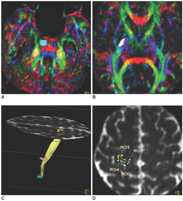
Fig. 1 Region of interest selection for diffusion tensor tractography at pons, posterior limb of internal capsule and precentral gyrus. A. Region of interest for pons was selected at expected corticopinal tract area on diffusion tensor MR imaging color map. Left side is left. B. Region of interest for corticospinal tract was selecteed at lateral half of posterior limb of internal capsule on diffusion tensor MR imaging color map. C. Precentral gyrus was determined by fibers crossing pons and posterior limb of internal capsule regions of interest, and it was identified by using anatomical landmarks such as posterior knob and superior frontal and precentral sulci on a B0 image. D. Four precentral gyrus regions of interest were drawn from medial (ROI 1) to lateral precentral gyrus (ROI 4) on B0 image. ROI 3 and ROI 4 were drawn near to posterior knob of precentral gyrus.

Fig. 2 Fiber tracts from medial half of posterior limb of internal capsule by single region of interest tracking. A-C. Fibers from medial posterior limb of internal capsule (purple color) descended to midbrain, which is posterior to cerebral peduncle. Fibers from medial posterior limb of internal capsule terminated at medial portion of precentral gyrus (medial to precentral gyrus regions of interest for corticospinal tracts), postcentral gyrus, and supplementary motor area.
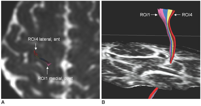
Fig. 3 Typical pattern of somatotopic organization of corticospinal tracts at posterior limb of internal capsule. A. Fiber tracts from lateral two regions of interest were located anterior to fiber tracts from medial two regions of interest at precentral gyrus as seen on B0 image (ROI 1-pink, ROI 2-blue, ROI 3-yellow, ROI 4-red). B. Corticospinal tract fibers were organized mainly along long axis of posterior limb of internal capsule. Medial fibers (ROI 1 and ROI 2, presumed foot fibers) were located more posterior to lateral fibers (ROI 3 and ROI 4, presumed hand fibers).
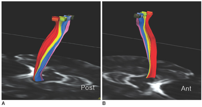
Fig. 4 Typical pattern of somatotopic organization of corticospinal tracts at cerebral peduncle. A, B. Corticospinal tract fibers were organized mainly along horizontal (left-to-right) axis of cerebral peduncle. Medial fibers (ROI 1 and ROI 2, presumed foot fibers) were located more lateral to lateral fibers (ROI 3 and ROI 4, presumed hand fibers).

Fig. 5 Intermixing of fibers from lateral regions of interest. A. ROI 3 fibers divided anterior and posterior division at precentral gyrus (B0 image). B, C. ROI 4 fibers were located between anterior and posterior divisions at posterior limb of internal capsule and cerebral peduncle.
Cited by 1 articles
-
Somatotopic Arrangement and Location of the Corticospinal Tract in the Brainstem of the Human Brain
Sung Ho Jang
Yonsei Med J. 2011;52(4):553-557. doi: 10.3349/ymj.2011.52.4.553.
Reference
-
1. Holodny AI, Gor DM, Watts R, Gutin PH, Ulug AM. Diffusion-tensor MR tractography of somatotopic organization of corticospinal tracts in the internal capsule: initial anatomic results in contradistinction to prior reports. Radiology. 2005. 234:649–653.2. Mori S, van Zijl PC. Fiber tracking: Principles and strategies - a technical review. NMR Biomed. 2002. 15:468–480.3. Wakana S, Jiang H, Nagae-Poetscher LM, van Zijl PC, Mori S. Fiber tract-based atlas of human white matter anatomy. Radiology. 2004. 230:77–87.4. Lee JS, Han MK, Kim SH, Kwon OK, Kim JH. Fiber tracking by diffusion tensor imaging in corticospinal tract stroke: Topographical correlation with clinical symptoms. Neuroimage. 2005. 26:771–776.5. Yamada K, Nagakane Y, Yoshikawa K, Kizu O, Ito H, Kubota T, et al. Somatotopic organization of thalamocortical projection fibers as assessed with MR tractography. Radiology. 2007. 242:840–845.6. Kido DK, LeMay M, Levinson AW, Benson WE. Computed tomographic localization of the precentral gyrus. Radiology. 1980. 135:373–377.7. Yousry TA, Schmid UD, Alkadhi H, Schmidt D, Peraud A, Buettner A, et al. Localization of the motor hand area to a knob on the precentral gyrus. A new landmark. Brain. 1997. 120:141–157.8. Kim JS, Pope A. Somatotopically located motor fibers in corona radiata: evidence from subcortical small infarcts. Neurology. 2005. 64:1438–1440.9. Newton JM, Ward NS, Parker GJ, Deichmann R, Alexander DC, Friston KJ, et al. Non-invasive mapping of corticofugal fibres from multiple motor areas-relevance to stroke recovery. Brain. 2006. 129:1844–1858.10. Behrens TE, Johansen-Berg H, Woolrich MW, Smith SM, Wheeler-Kingshott CA, Boulby PA, et al. Non-invasive mapping of connections between human thalamus and cortex using diffusion imaging. Nat Neurosci. 2003. 6:750–757.11. Parker GJ, Alexander DC. Probabilistic Monte Carlo based mapping of cerebral connections utilizing whole-brain crossing fibre information. Inf Process Med Imaging. 2003. 18:684–695.12. Parker GJ, Haroon HA, Wheeler-Kingshott CA. A framework for a streamline-based probabilistic index of connectivity (PICo) using a structural interpretation of MRI diffusion measurements. J Magn Reson Imaging. 2003. 18:242–254.13. Holodny AI, Watts R, Korneinko VN, Pronin IN, Zhukovskiy ME, Gor DM, et al. Diffusion tensor tractography of the motor white matter tracts in man: Current controversies and future directions. Ann N Y Acad Sci. 2005. 1064:88–97.
- Full Text Links
-
- Actions
-
Cited
- CITED
-
- Close
- Share
-
- Similar articles
-
- Assessment of Normal-Appearing White Matter Damage in Multiple Sclerosis Using Diffusion Tensor Imaging
- Diffusion Tensor Tractography in Two Cases of Kernohan-Woltman Notch Phenomenon
- Usefulness of DTI-based three dimensional corticospinal tractography in children with hemiplegic cerebral palsy
- Somatotopic Arrangement and Location of the Corticospinal Tract in the Brainstem of the Human Brain
- Applicability of Diffusion Tensor MR Imaging and Fiber Tractography That Predict Short-Term Functional Motor Outcome in Patients with Deep Intracerebral Hemorrhage
