J Korean Surg Soc.
2010 Apr;78(4):238-241. 10.4174/jkss.2010.78.4.238.
Clinical Experiences of Pilimatrixoma in a Single Institution
- Affiliations
-
- 1Department of Surgery, Gangnam Severance Hospital, Yonsei University College of Medicine, Seoul, Korea. medilys@yuhs.ac
- 2Department of Plastic and Reconstructive Surgery, Gangnam Severance Hospital, Yonsei University College of Medicine, Seoul, Korea.
- 3Department of Pathology, Gangnam Severance Hospital, Yonsei University College of Medicine, Seoul, Korea.
- KMID: 1750736
- DOI: http://doi.org/10.4174/jkss.2010.78.4.238
Abstract
- PURPOSE
To describe the clinical presentations, management, and outcomes of patients with pilomatrixomas treated in a single institution, and to compare the clinicopathological features according to their location.
METHODS
We reviewed the medical records of 57 patients treated between January 1986 and December 2007, retrospectively.
RESULTS
The 57 patients had a total of 61 cases of pilomatrixomas. The mean age at diagnosis was 12.4 years, with most patients aged 0~10 years, followed by 10~20 years and 20~30 years. Mean tumor size was 1.46 cm, and most tumors were less than 2.0 cm in diameter. Forty-two tumors (68.9%) on the head and the neck were classified as Group I, and 19 tumors (31.1%) on the body were classified as Group II. The mean age at diagnosis was 9.4 years in Group I, and 19.8 years in Group II with significant statistical difference (P=0.009). The mean duration of symptoms was 26.4 months in Group I, and 7.2 months in Group II (P=0.001). All patients were treated surgically, and two patients relapsed (5 months and 3 years later).
CONCLUSION
Pilomatrixoma is an uncommon benign skin tumor arising from hair follicle matrix cells. Diagnosis is usually easy based on clinical findings, and preoperative diagnosis may be improved with increased awareness of pilomatrixoma. Complete surgical excision is the treatment of choice, and recurrence after complete excision is rare.
Keyword
Figure
-
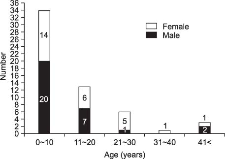
Fig. 1 Age and gender distribution of patients (n=57).
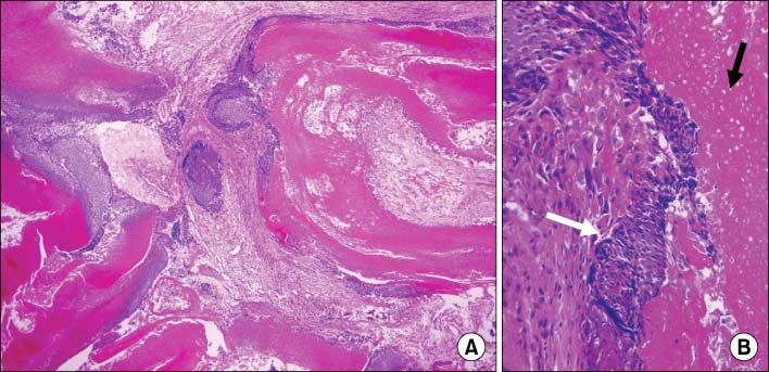
Fig. 2 Histopathologic findings of pilomatrixoma. (A) At lower power view, it is composed of several cystic structures with keratin lake (H&E, ×40). (B) In high power view, the cystic space is lined by basaloid squamous cells (white arrow) and ghost cells (black arrow) (H&E, ×200).
Reference
-
1. Malherbe A, Chenantais J. Note sur l'epithelioma calcifiedes glandes sebacees. Prog Med. 1880. 8:826–837.2. Lan MY, Lan MC, Ho CY, Li WY, Lin CZ. Pilomatricoma of the head and neck: a retrospective review of 179 cases. Arch Otolaryngol Head Neck Surg. 2003. 129:1327–1330.3. Greene RM, McGuff HS, Miller FR. Pilomatrixoma of the face: a benign skin appendage mimicking squamous cell carcinoma. Otolaryngol Head Neck Surg. 2004. 130:483–485.4. Moehlenbeck FW. Pilomatrixoma (calcifying epithelioma). A statistical study. Arch Dermatol. 1973. 108:532–534.5. Aslan G, Erdogan B, Akoz T, Gorgu M, Seckin S, Terzioglu A. Multiple occurrence of pilomatrixoma. Plast Reconstr Surg. 1996. 98:510–513.6. Singh B, Tolete-Velcek F, Alexis R. Pathological case of the month. Pilomatrixoma. Arch Pediatr Adolesc Med. 1995. 149:551–552.7. McBrien M, Victor T, Wolff AP. Pathologic quiz case 2. Pilomatrixoma. Arch Otolaryngol Head Neck Surg. 1988. 114:1042–1043. 10458. Aissoudi M, Doss N, Bouzaiene A. A case for diagnosis: pilomatrixoma. Ann Dermatol Venereol. 1995. 122:797–798.9. Hawkins DB, Chen WT. Pilomatrixoma of the head and neck in children. Int J Pediatr Otorhinolaryngol. 1985. 8:215–223.10. Urvoy M, Legall F, Toulemont PJ, Chevrant-Breton J. Multiple pilomatricoma. Apropos of a case. J Fr Ophtalmol. 1996. 19:464–466.11. Duflo S, Nicollas R, Roman S, Magalon G, Triglia JM. Pilomatrixoma of the head and neck in children: a study of 38 cases and a review of the literature. Arch Otolaryngol Head Neck Surg. 1998. 124:1239–1242.12. Brami S, Riquet C, Cattan P, Spinel W. A propos de nodules jugaux: une entite remarquable: l'epithelioma calcifie de Malherbe. Cahiers ORL. 1980. 15:65–67.13. Domanski HA, Domanski AM. Cytology of pilomatrixoma (calcifying epithelioma of Malherbe) in fine needle aspirates. Acta Cytol. 1997. 41:771–777.14. Phyu KK, Bradley PJ. Pilomatrixoma in the parotid region. J Laryngol Otol. 2001. 115:1026–1028.15. Saussez S, Mahillon V, Blaivie C, Haller A, Chantrain G, Thill MP. Aggressive pilomatrixoma of the infra-auricular area: a case report. Auris Nasus Larynx. 2005. 32:407–410.
- Full Text Links
-
- Actions
-
Cited
- CITED
-
- Close
- Share
-
- Similar articles
-
- Techniques of gynecologic single-port access laparoscopic surgery
- Single-Incision Video-Assisted Thoracoscopic Surgery for Benign Mediastinal Diseases: Experiences in Single Institution
- Career choice experiences of nursing students
- Focal therapy for prostate cancer with irreversible electroporation: Oncological and functional results of a single institution study
- Experiences of Nurses Working in a Single-Room-Structured Intensive Care Unit
