Interstitial Injection Mammoplasty Mimicking Diffuse Infiltrative Disease in a Male Patient
- Affiliations
-
- 1Department of Radiology, Ewha Womans University School of Medicine, Mokdong Hospital, Seoul, Korea. escha@ewha.ac.kr
- 2Department of Pathology, Ewha Womans University School of Medicine, Mokdong Hospital, Seoul, Korea.
- KMID: 1748456
- DOI: http://doi.org/10.3348/jksr.2013.68.3.225
Abstract
- Foreign material injection for breast augmentation has been performed for decades, primarily in Asia. Various materials have been used clinically for injection; their typical imaging findings are well-known and have been reported in many cases. However, these cases usually involve an injection of a foreign material for female breast augmentation. We report here the unusual imaging findings in a 64-year-old male with lipogranulomatous inflammatory changes in his breast, caused by an interstitial injection of paraffin. Mammograms show the enlargement of both breasts as well as an increased density with combined skin and trabecular thickening. Ultrasonography revealed bilateral, severely thickened skin and diffuse edematous change along the subcutaneous fat layer. Further, a few oil cysts and ill-defined hypoechoic lesions with posterior acoustic shadowing were noted in both breasts.
MeSH Terms
Figure
-
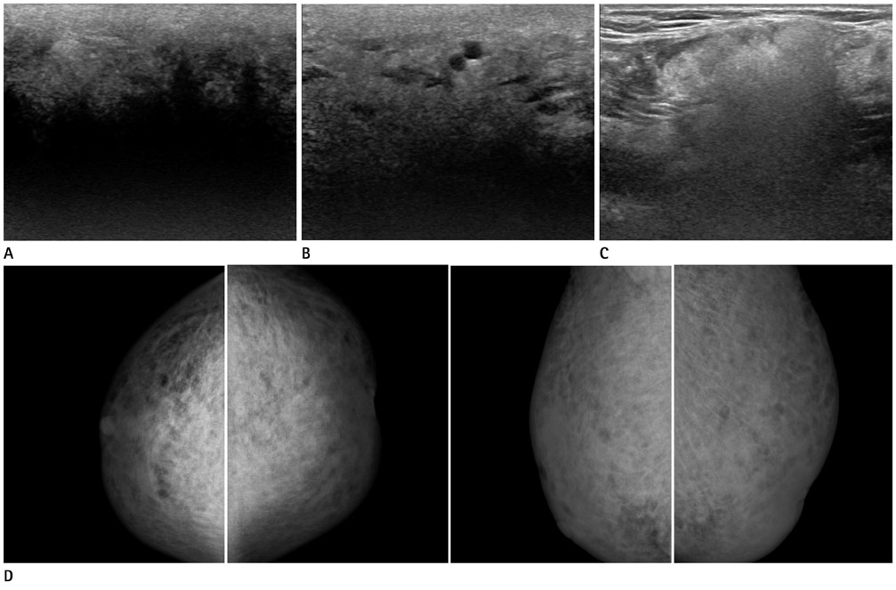
Fig. 1 A 64-year-old male patient with breast enlargement. Breast ultrasound showed diffuse edematous changes along the subcutaneous fat layer and surface skin thickening in both breasts. A. Diffuse intraparenchymal shadowing similar to the "snowstorm" appearance was seen in the center of both breasts. B. Round-to-oval anechoic cysts, suggestive of oil cysts, were seen in both breasts. C. Paraffin uptake by lymph nodes was seen as diffuse increased echogenic lesions with dirty acoustic shadowing in both axillae. D. Bilateral standard mammograms showed enlargement of both breasts and increased density with combined skin and trabecular thickening.

Fig. 2 Follow-up sonography 14 months later. A. Breast ultrasonography showed ill-defined hypoechoic lesions with diffuse intraparenchymal shadowing in both breasts without interval change. B. Ultrasound-guided core needle biopsy using a 14-gauge needle was performed in the upper portion of both breasts. C. Photomicrograph showed foreign body granuloma with severe sclerosing lipogranulomatous inflammation, compatible with paraffinoma (hematoxylin & eosin; × 200).
Reference
-
1. Khedher NB, David J, Trop I, Drouin S, Peloquin L, Lalonde L. Imaging findings of breast augmentation with injected hydrophilic polyacrylamide gel: patient reports and literature review. Eur J Radiol. 2011. 78:104–111.2. Yang N, Muradali D. The augmented breast: a pictorial review of the abnormal and unusual. AJR Am J Roentgenol. 2011. 196:W451–W460.3. Youk JH, Son EJ, Kim EK, Kim JA, Kim MJ, Kwak JY, et al. Diagnosis of breast cancer at dynamic MRI in patients with breast augmentation by paraffin or silicone injection. Clin Radiol. 2009. 64:1175–1180.4. Han BK, Choe YH, Ko YH, Nam SJ, Yang JH. Foreign body granulomas of the breast presenting as bilateral spiculated masses. Korean J Radiol. 2001. 2:113–116.5. Teo SY, Wang SC. Radiologic features of polyacrylamide gel mammoplasty. AJR Am J Roentgenol. 2008. 191:W89–W95.6. Irshad A, Ackerman SJ, Pope TL, Moses CK, Rumboldt T, Panzegrau B. Rare breast lesions: correlation of imaging and histologic features with WHO classification. Radiographics. 2008. 28:1399–1414.7. Sabaté JM, Clotet M, Gómez A, De Las Heras P, Torrubia S, Salinas T. Radiologic evaluation of uncommon inflammatory and reactive breast disorders. Radiographics. 2005. 25:411–424.8. Iyengar R, Saint-Cyr M, Gokaslan T, Rao R. Breast paraffinoma. Breast J. 2008. 14:504–505.9. Markopoulos C, Mantas D, Kouskos E, Antonopoulou Z, Revenas C, Yiacoumettis A. Paraffinomas of the breast or oleogranulomatous mastitis-a rare entity. Breast. 2006. 15:540–543.
- Full Text Links
-
- Actions
-
Cited
- CITED
-
- Close
- Share
-
- Similar articles
-
- Diffuse Interstitial Infiltrative Lung Metastasis of Malignant Melanoma: a Case Report
- DILD (diffuse infiltrative lung disease); Radiologic Diagnostic Approach According to High-Resolution CT Pattern
- MR Findings of Siliconoma in Interstitial Silicone Injection Mammoplasty Patients
- A Case of Diffuse Infiltrative Lymphocytosis Syndrome Associated with Human Immunodeficiency Virus Infection
- The Value of MRI Findings in Augmented Mammoplasty
