J Vet Sci.
2014 Mar;15(1):149-155. 10.4142/jvs.2014.15.1.149.
Echocardiographic assessment of coronary artery flow in normal canines and model dogs with myocardial infarction
- Affiliations
-
- 1Department of Veterinary Radiology and Diagnostic Imaging, College of Veterinary Medicine, Konkuk University, Seoul 143-701, Korea. eomkd@konkuk.ac.kr
- KMID: 1737621
- DOI: http://doi.org/10.4142/jvs.2014.15.1.149
Abstract
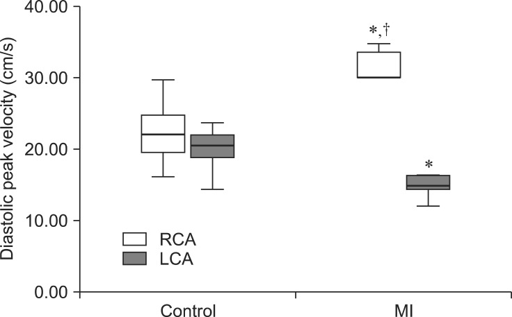
- This study was conducted to evaluate the usefulness of coronary arterial profiles from normal dogs (11 animals) and canines (six dogs) with experimental myocardial infarction (MI) induced by ligation of the left coronary artery (LCA). Blood velocity of the LCA and right coronary artery (RCA) were evaluated following transthoracic pulsed-wave Doppler echocardiography. The LCA was observed as an infundibular shape, located adjacent to the sinus of Valsalva. The RCA appeared as a tubular structure located 12 o'clock relative to the aorta. In normal dogs, the LCA and RCA mean peak diastolic velocities were 20.84 +/- 3.24 and 19.47 +/- 2.67 cm/sec, respectively. The LCA and RCA mean diastolic deceleration times were 0.91 +/- 0.14 sec and 1.13 +/- 0.20 sec, respectively. In dogs with MI, the LCA had significantly (p < 0.01) lower peak velocities (14.82 +/- 1.61 cm/sec) than the RCA (31.61 +/- 2.34 cm/sec). The RCA had a significantly (p < 0.01) rapid diastolic deceleration time (0.71 +/- 0.06 sec) than that found in the LCA (1.02 +/- 0.22 sec) of MI dogs. In conclusion, these profiles may serve as a differential factor for evaluating cardiomyopathy in dogs.
MeSH Terms
Figure
Reference
-
1. Akasaka T, Yoshida K, Hozumi T, Takagi T, Kaji S, Kawamoto T, Ueda Y, Okada Y, Morioka S, Yoshikawa J. Restricted coronary flow reserve in patients with mitral regurgitation improves after mitral reconstructive surgery. J Am Coll Cardiol. 1998; 32:1923–1930. PMID: 9857873.2. Bossbaly MJ, Buchanan JW, Sammaro C. Aortic body carcinoma and myocardial infarction in a dobermann pinscher. J Small Anim Pract. 1993; 34:638–642.
Article3. Botnar RM, Stuber M, Danias PG, Kissinger KV, Manning WJ. Improved coronary artery definition with T2-weighted, free-breathing, three-dimensional coronary MRA. Circulation. 1999; 99:3139–3148. PMID: 10377077.
Article4. Buchanan JW. Pathogenesis of single right coronary artery and pulmonic stenosis in English bulldogs. J Vet Intern Med. 2001; 15:101–104. PMID: 11300591.
Article5. Carabello BA, Nakano K, Ishihara K, Kanazawa S, Biederman RWW, Spann JF Jr. Coronary blood flow in dogs with contractile dysfunction due to experimental volume overload. Circulation. 1991; 83:1063–1075. PMID: 1825623.
Article6. Chansky M, Levy MN. Collateral circulation to myocardial regions supplied by anterior descending and right coronary arteries in the dog. Circ Res. 1962; 11:414–417. PMID: 14020098.
Article7. Connolly DJ, Cannata J, Boswood A, Archer A, Groves EA, Neiger R. Cardiac troponin I in cats with hypertrophic cardiomyopathy. J Feline Med Surg. 2003; 5:209–216. PMID: 12878148.
Article8. Cunningham SM, Rush JE, Freeman LM. Systemic inflammation and endothelial dysfunction in dogs with congestive heart failure. J Vet Intern Med. 2012; 26:547–557. PMID: 22489997.
Article9. Egenvall A, Bonnett BN, Häggström J. Heart disease as a cause of death in insured Swedish dogs younger than 10 years of age. J Vet Intern Med. 2006; 20:894–903. PMID: 16955814.
Article10. Falk T, Jönsson L, Olsen LH, Pedersen HD. Arteriosclerotic changes in the myocardium, lung, and kidney in dogs with chronic congestive heart failure and myxomatous mitral valve disease. Cardiovasc Pathol. 2006; 15:185–193. PMID: 16844549.
Article11. Falk T, Jönsson L, Pedersen HD. Intramyocardial arterial narrowing in dogs with sub aortic stenosis. J Small Anim Pract. 2004; 45:448–453. PMID: 15460203.12. Fox PR. Hypertrophic cardiomyopathy. Clinical and pathological correlates. J Vet Cardiol. 2003; 5:39–45. PMID: 19081364.13. Haider B, Ahmed SS, Moschos CB, Oldewurtel HA, Regan TJ. Myocardial function and coronary blood flow response to acute ischemia in chronic canine diabetes. Circ Res. 1977; 40:577–583. PMID: 870238.
Article14. Hernandez JL, Bélanger MC, Benoit-Biancamano MO, Girard C, Pibarot P. Left coronary aneurysmal dilation and subaortic stenosis in a dog. J Vet Cardiol. 2008; 10:75–79. PMID: 18485856.
Article15. Hess RS, Kass PH, Van Winkle TJ. Association between diabetes mellitus, hypothyroidism or hyperadrenocorticism, and atherosclerosis in dogs. J Vet Intern Med. 2003; 17:489–494. PMID: 12892299.
Article16. Heusch G, Deussen A. The effects of cardiac sympathetic nerve stimulation on perfusion of stenotic coronary arteries in the dog. Circ Res. 1983; 53:8–15. PMID: 6861299.
Article17. Hozumi T, Yoshida K, Akasaka T, Asami Y, Ogata Y, Takagi T, Kaji S, Kawamoto T, Ueda Y, Morioka S. Noninvasive assessment of coronary flow velocity and coronary flow velocity reserve in the left anterior descending coronary artery by Doppler echocardiography: comparison with invasive technique. J Am Coll Cardiol. 1998; 32:1251–1259. PMID: 9809933.18. Kingma JG Jr, Vincent C, Rouleau JR, Kingma I. Influence of acute renal failure on coronary vasoregulation in dogs. J Am Soc Nephrol. 2006; 17:1316–1324. PMID: 16597686.
Article19. Kittleson MD, Meurs KM, Munro MJ, Kittleson JA, Liu SK, Pion PD, Towbin JA. Familial hypertrophic cardiomyopathy in maine coon cats: an animal model of human disease. Circulation. 1999; 99:3172–3180. PMID: 10377082.20. Kurita T, Sakuma H, Onishi K, Ishida M, Kitagawa K, Yamanaka T, Tanigawa T, Kitamura T, Takeda K, Ito M. Regional myocardial perfusion reserve determined using myocardial perfusion magnetic resonance imaging showed a direct correlation with coronary flow velocity reserve by Doppler flow wire. Eur Heart J. 2009; 30:444–452. PMID: 19098020.
Article21. Moesgaard SG, Pederson LG, Teerlink T, Häggström J, Pederson HD. Neurohormonal and circulatory effects of short-term treatment with enalapril and quinapril in dogs with asymptomatic mitral regurgitation. J Vet Intern Med. 2005; 19:712–719. PMID: 16231716.
Article22. Pannu HK, Flohr TG, Corl FM, Fishman EK. Current concepts in multi-detector row CT evaluation of the coronary arteries: principles, techniques, and anatomy. Radiographics. 2003; 23(Suppl 1):S111–S125. PMID: 14557506.
Article23. Pyle RL, Lowensohn HS, Khouri EM, Gregg DE, Patterson DF. Left circumflex coronary artery hemodynamics in conscious dogs with congenital subaortic stenosis. Circ Res. 1973; 33:34–38. PMID: 4271850.
Article24. Ropers D, Baum U, Pohle K, Anders K, Ulzheimer S, Ohnesorge B, Schlundt C, Bautz W, Daniel WG, Achenbach S. Detection of coronary artery stenoses with thin-slice multi-detector row spiral computed tomography and multiplanar reconstruction. Circulation. 2003; 107:664–666. PMID: 12578863.
Article25. Scheffel H, Stolzmann P, Plass A, Leschka S, Grünenfelder J, Falk V, Marincek B, Alkadhi H. Coronary artery disease in patients with cardiac tumors: preoperative assessment by computed tomography coronary angiography. Interact Cardiovasc Thorac Surg. 2010; 10:513–518. PMID: 20118120.
Article26. Scott JC, Balourdas TA, Croll MN. The effect of experimental hypothyroidism on coronary blood flow and hemodynamic factors. Am J Cardiol. 1961; 7:690–693. PMID: 13749361.
Article27. Shannon RP, Stambler BS, Komamura K, Ihara T, Vatner SF. Cholinergic modulation of the coronary vasoconstriction induced by cocaine in conscious dogs. Circulation. 1993; 87:939–949. PMID: 8443913.
Article28. Tanaka R, Murota A, Nagashima Y, Yamane Y. Changes in Platelet life span in dogs with mitral valve regurgitation. J Vet Intern Med. 2002; 16:446–451. PMID: 12141307.
Article29. Tarnow I, Kristensen AT, Olsen LH, Falk T, Haubro L, Pedersen LG, Pedersen HD. Dogs with heart disease causing turbulent high-velocity blood flow have changes in platelet function and von Willebrand factor multimer distribution. J Vet Intern Med. 2005; 19:515–522. PMID: 16095168.30. Teragaki M, Toda I, Takagi M, Fukuda S, Ujino K, Takeuchi K, Yoshikawa J. New applications of intracardiac echocardiography: assessment of coronary blood flow by colour and pulsed Doppler imaging in dogs. Heart. 2002; 88:283–288. PMID: 12181224.
Article31. Tune JD, Yeh C, Setty S, Zong P, Downey HF. Coronary blood flow control is impaired at rest and during exercise in conscious diabetic dogs. Basic Res Cardiol. 2002; 97:248–257. PMID: 12061395.
Article32. Vollmar A, Fox PR, Meurs KM, Liu SK. Dilated cardiomyopathy in juvenile Doberman pinschers. J Vet Cardiol. 2003; 5:23–27. PMID: 19081354.
Article
- Full Text Links
- Actions
-
Cited
- CITED
-
- Close
- Share
- Similar articles
-
- Coronary Slow Flow Phenomenon Leads to ST Elevation Myocardial Infarction
- A Case of Non-Q Myocardial Infaction in a Patient with Myocardial Bridging
- Effects of Myocardial Stunning on Remote Coronary Flow Reserve
- Coronary-Pulmonary Fistulas Involving All Three Major Coronary Arteries Co-Existing With Myocardial Infarction
- Assessment of Myocardial Collateral Blood Flow with Contrast Echocardiography


