A Case of Eosinophilic Fasciitis: Imaging Findings from Early Diagnosis to Complete Remission
- Affiliations
-
- 1Department of Radiology, Dankook University Hospital, Cheonan, Korea. jeelee_0@naver.com
- KMID: 1736482
- DOI: http://doi.org/10.3348/jksr.2014.71.3.150
Abstract
- Eosinophilic fasciitis (EF) is a rare disease characterized by symmetric and painful swelling with progressive induration and thickening of the skin and soft tissues of the limbs and trunk. Herein, we report a case of a 31-year-old woman who presented with painful swelling in both lower legs which persisted for 6 days. She underwent ultrasonography (US) in an out-patient department to rule out deep-vein thrombosis. The US did, however, reveal perifascial fluid in the thickened superficial fascia and interstitial fluid in the subcutaneous layer of both lower legs. Magnetic resonance imaging findings were identical to the US and additionally showed no involvement of the muscles or deep fascia. Laboratory data, showing peripheral eosinophilia and a US-guided gun-biopsy showing lymphocytic and eosinophilic infiltration were both indicative of EF. The patient was treated with corticosteroids, resulting in a remarkable improvement in both the lower-leg edema and peripheral eosinophilia. There was no recurrence after 7 years of follow-up.
MeSH Terms
Figure
-
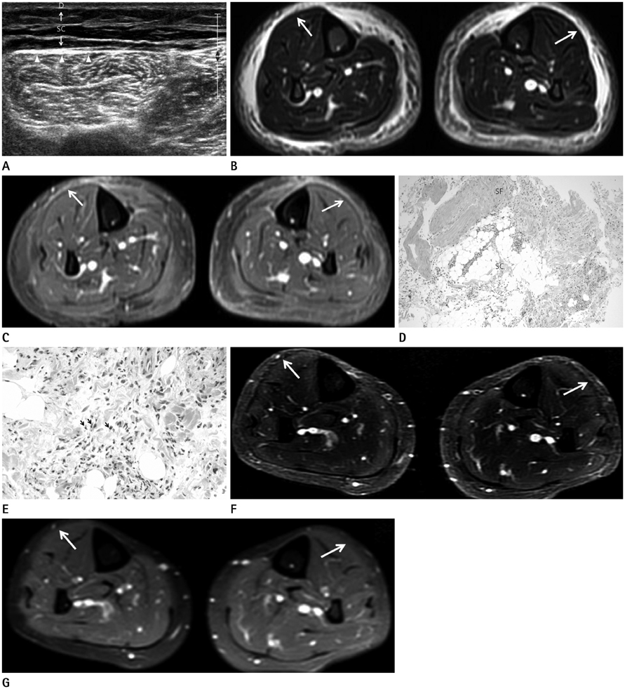
Fig. 1 Eosinophilic fasciitis (EF) in both lower legs of 31-year-old woman. A. Ultrasonography (US) of right-lower leg shows diffuse interstitial fluid in thickened subcutaneous layer (space between arrows) and superficial fascia (arrowheads). There is no intramuscular focal lesion or perifascial fluid in the deep fascia, nor is there any evidence of deep-vein thrombosis (not shown). B, C. Magnetic resonance imaging (MRI) of both lower leg shows interstitial fluid (B, arrows) in subcutaneous layer and superficial fascia on fat-suppressed T2-weighted image (B) and subtle linear enhancement (C, arrows) on fat-suppressed contrast-enhanced T1-weighted image (C). There is no signal change or enhancement within the muscle or deep fascia. D. Photomicrograph (H&E stain, × 100) of fascia-subcutaneous fat junction showing thickened fascia and entrapment of subcutaneous fat by heavy inflammatory cell infiltration. E. High-power photomicrograph (H&E stain, × 400) of fascia showing heavy inflammatory cell infiltration with numerous eosinophils (arrows), lymphocytes and occasional plasma cells. F, G. MRI was obtained after 6 months of corticosteroids treatment. Fat-suppressed T2-weighted image (F) and fat-suppressed contrast-enhanced T1-weighted image (G) showing complete remission. The patient is symptom-free, and the laboratory findings are normal. The arrows show the sites of abnormalities identified in a previous MRI. Note.-D = dermal layer, SC = subcutaneous layer, SF = superficial fascia
Reference
-
1. Shulman LE. Diffuse fasciitis with hypergammaglobulinemia and eosinophilia: a new syndrome? J Rheumatol. 1984; 11:569–570.2. Pinal-Fernandez I, Selva-O'Callaghan A, Grau JM. Diagnosis and classification of eosinophilic fasciitis. Autoimmun Rev. 2014; 13:379–382.3. Lakhanpal S, Ginsburg WW, Michet CJ, Doyle JA, Moore SB. Eosinophilic fasciitis: clinical spectrum and therapeutic response in 52 cases. Semin Arthritis Rheum. 1988; 17:221–231.4. Barnes L, Rodnan GP, Medsger TA, Short D. Eosinophilic fasciitis. A pathologic study of twenty cases. Am J Pathol. 1979; 96:493–451.5. Moulton SJ, Kransdorf MJ, Ginsburg WW, Abril A, Persellin S. Eosinophilic fasciitis: spectrum of MRI findings. AJR Am J Roentgenol. 2005; 184:975–978.6. Baumann F, Brühlmann P, Andreisek G, Michel BA, Marincek B, Weishaupt D. MRI for diagnosis and monitoring of patients with eosinophilic fasciitis. AJR Am J Roentgenol. 2005; 184:169–174.7. Desvignes-Engelbert A, Saulière N, Loeuille D, Blum A, Chary-Valckenaere I. From diagnosis to remission: place of MRI in eosinophilic fasciitis. Clin Rheumatol. 2010; 29:1461–1464.8. Lee HS, Chang SJ, Kang MS, Yoon CS, Kim KW, Sohn MH, et al. A case of eosinophilic fasciitis presenting as pitting edema of the lower extremities. Allergy Asthma Immunol Res. 2014; 6:179–182.9. Chan V, Soans B, Mathers D. Ultrasound and magnetic resonance imaging features in a patient with eosinophilic fasciitis. Australas Radiol. 2004; 48:414–417.
- Full Text Links
-
- Actions
-
Cited
- CITED
-
- Close
- Share
-
- Similar articles
-
- Eosinophilic Fasciitis Localized on the Left Lower Extremity: A case report
- Eosinophilic Fasciitis Associated with Overlying Intraepidermal Blister Formation: A Case Report
- Eosinophilic Fasciitis and Musculoskeletal Complication in a Child : A case report
- A Case of Eosinophilic Fasciitis
- A Case of Eosinophilic Fasciitis
