Tissue Responses to Stent Grafts with Endo-Exo-Skeleton for Saccular Abdominal Aortic Aneurysms in a Canine Model
- Affiliations
-
- 1Department of Radiology and Institute of Radiation Medicine, Seoul National University College of Medicine, Clinical Research Institute, Seoul 110-744, Korea.
- 2Department of Radiology, Seoul National University Boramae Hospital, Seoul 156-707, Korea. cyho50168@naver.com
- 3Department of Radiology, National Cancer Center, Goyang 410-769, Korea.
- 4Department of Surgery, Seoul National University College of Medicine, Seoul 110-744, Korea.
- 5Department of Radiology, Gachon University Gil Medical Center, Incheon 405-760, Korea.
- KMID: 1734947
- DOI: http://doi.org/10.3348/kjr.2014.15.5.622
Abstract
OBJECTIVE
We evaluated the effect of close contact between the stent and the graft on the induction of endothelial covering on the stent graft placed over an aneurysm.
MATERIALS AND METHODS
Saccular abdominal aortic aneurysms were made with Dacron patch in eight dogs. The stent graft consisted of an inner stent, a expanded polytetrafluoroethylene graft, and an outer stent. After sacrificing the animals, the aortas with an embedded stent graft were excised. The aortas were inspected grossly and evaluated microscopically.
RESULTS
The animals were sacrificed at two (n = 3), six (n = 3), and eight months (n = 2) after endovascular repair. In two dogs, the aortic lumen was occluded at two months after the placement. On gross inspection of specimens from the other six dogs with a patent aortic lumen, stent grafts placed over the normal aortic wall were covered by glossy white neointima, whereas, stent grafts placed over the aneurysmal aortic wall were covered by brownish neointima. On microscopic inspection, stent grafts placed over the normal aortic wall were covered by thin neointima (0.27 +/- 0.05 mm, mean +/- standard deviation) with an endothelial layer, and stent grafts placed over the aneurysmal aortic wall were covered by thick neointima (0.62 +/- 0.17 mm) without any endothelial lining. Transgraft cell migration at the normal aortic wall was more active than that at the aneurysmal aortic wall.
CONCLUSION
Close contact between the stent and the graft, which was achieved with stent grafts with endo-exo-skeleton, could not enhance endothelial covering on the stent graft placed over the aneurysms.
Keyword
MeSH Terms
Figure
-

Fig. 1 Stent graft with endo-exo-skeleton consisting of inner bare stent, middle graft, and outer bare stent.
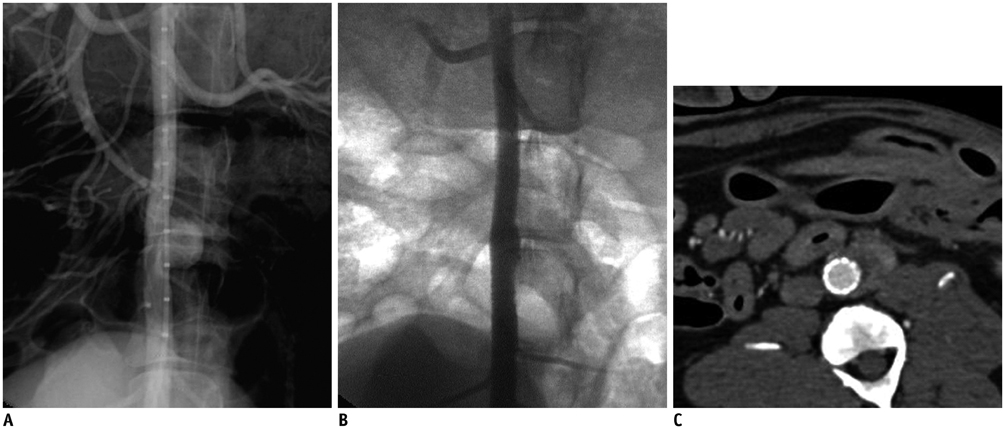
Fig. 2 Angiography before and after stent graft placement. A. Conventional angiography shows aortic saccular aneurysm to left of aorta. B. On completion angiography after stent graft placement, middle part of inserted stent graft shows outward convexity towards saccular aneurysm. C. CT angiography at two months demonstrates complete exclusion of aneurysm, no endoleak, and patent lumen.
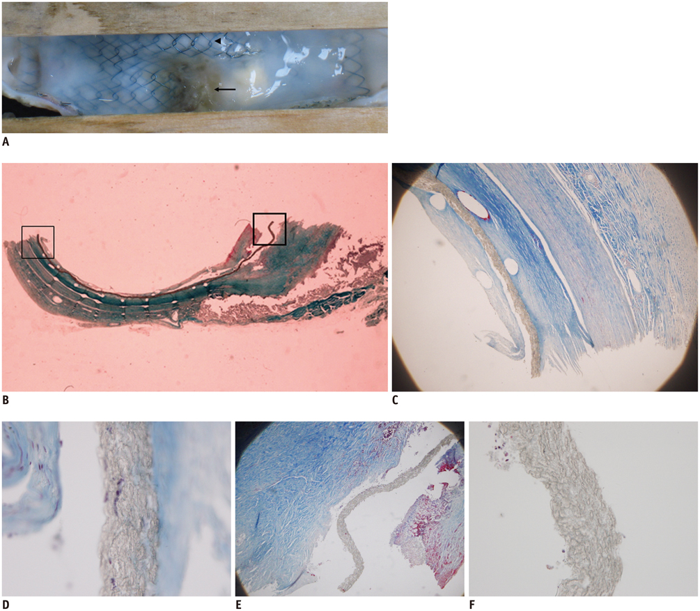
Fig. 3 Gross inspection findings and transgraft cell migration. A. Gross inspection after longitudinal incision of abdominal aorta at opposite side of aneurysm demonstrates thick brown neointima (arrow) over aneurysmal aortic wall, which protrudes towards aortic lumen. Glossy white neointima (arrowhead) is noted over normal aortic wall. B. Microscopic examination (Masson's trichrome stain, original magnification × 1) shows thin neointima over normal aortic wall (thin-lined rectangular block) and thick neointima over aneurysmal aortic wall (thick-lined rectangular block). C, D. Magnified view of thin-lined rectangular block in B located in normal aortic wall (original magnification, × 40 and × 400) shows graft with lot of collagen (bright blue) and more than ten spindle cells (violet) rated as Grade 3. Brown band between footprints of inner and outer stent wire struts in C and E is graft. E, F. Magnified view of thick-lined rectangular block in B located in stent graft placed over excluded saccular aneurysm (original magnification, × 40 and × 400) shows graft with little collagen and few spindle cells rated as Grade 1.

Fig. 4 Immunohistochemical staining of endothelial covering. A. Cross-section of bisected aorta contains stent graft and aneurysm (Factor VIII-related antigen immunohistochemical staining, original magnification × 1). Thin-lined rectangular block is normal aortic wall and thick-lined rectangular block is aneurysmal aortic wall. B, C. Magnified view of thin-lined rectangular block in A located in normal aorta (B; Factor VIII-related antigen immunohistochemical staining, C; Hematoxylin-Eosin staining, original magnification, × 400) shows layer of cells strongly reactive to Factor VIII-related antigen immunohistochemical staining, which indicates endothelial covering on neointima. D, E. Magnified views of thick-lined rectangular block in A located in stent graft placed over excluded saccular aneurysm (D; Factor VIII-related antigen immunohistochemical staining, E; Hematoxylin-Eosin staining, original magnification, × 400) show layer of cells in neointima that are not stained on Factor VIII-related antigen immunohistochemical staining.
Reference
-
1. Parodi JC, Palmaz JC, Barone HD. Transfemoral intraluminal graft implantation for abdominal aortic aneurysms. Ann Vasc Surg. 1991; 5:491–499.2. Noishiki Y, Tomizawa Y, Yamane Y, Matsumoto A. The vicious cycle of nonhealing neointima in fabric vascular prostheses. Artif Organs. 1995; 19:7–16.3. Berger K, Sauvage LR, Rao AM, Wood SJ. Healing of arterial prostheses in man: its incompleteness. Ann Surg. 1972; 175:118–127.4. Laborde JC, Parodi JC, Clem MF, Tio FO, Barone HD, Rivera FJ, et al. Intraluminal bypass of abdominal aortic aneurysm: feasibility study. Radiology. 1992; 184:185–190.5. Dolmatch BL, Tio FO, Li XD, Dong YH. Patency and tissue response related to two types of polytetrafluoroethylene-covered stents in the dog. J Vasc Interv Radiol. 1996; 7:641–649.6. Marty B, Leu AJ, Mucciolo A, von Segesser LK. Biologic fixation of polyester-versus polyurethane-covered stents in a porcine model. J Vasc Interv Radiol. 2002; 13:601–607.7. Wilson EP, White RA, Kopchok GE, Donayre CE, de Virgilio C, Geselschap JH, et al. Deployment and healing of an ePTFE encapsulated stent endograft in the canine aorta. Ann Vasc Surg. 1997; 11:354–358.8. Virmani R, Kolodgie FD, Dake MD, Silver JH, Jones RM, Jenkins M, et al. Histopathologic evaluation of an expanded polytetrafluoroethylene-nitinol stent endoprosthesis in canine iliofemoral arteries. J Vasc Interv Radiol. 1999; 10:445–456.9. Boudghène F, Anidjar S, Allaire E, Osborne-Pellegrin M, Bigot JM, Michel JB. Endovascular grafting in elastase-induced experimental aortic aneurysms in dogs: feasibility and preliminary results. J Vasc Interv Radiol. 1993; 4:497–504.10. Palmaz JC, Tio FO, Laborde JC, Clem M, Rivera FJ, Murphy KD, et al. Use of stents covered with polytetrafluoroethylene in experimental abdominal aortic aneurysm. J Vasc Interv Radiol. 1995; 6:879–885.11. Benson AE, Palmaz JC, Tio FO, Sprague EA, Encarnacion CE, Josephs SC. Polytetrafluoroethylene-encapsulated stent-grafts: use in experimental abdominal aortic aneurysm. J Vasc Interv Radiol. 1999; 10:605–612.12. Kim HB, Choi YH, So YH, Min SK, Kim HC, Kim YI, et al. Tissue responses to endovascular stent grafts for saccular abdominal aortic aneurysms in a canine model. J Korean Med Sci. 2012; 27:1170–1176.13. Makutani S, Kichikawa K, Uchida H, Maeda M, Konishi N, Hiasa Y, et al. Effect of antithrombotic agents on the patency of PTFE-covered stents in the inferior vena cava: an experimental study. Cardiovasc Intervent Radiol. 1999; 22:232–238.14. Ishii S, Sato M, Sonomura T, Yamada K, Tanihata H, Ishikawa H, et al. Optimal covering material for stent-grafts placed in the portal vein in a canine model. Cardiovasc Intervent Radiol. 2005; 28:624–631.15. Ombrellaro MP, Stevens SL, Kerstetter K, Freeman MB, Goldman MH. Healing characteristics of intraarterial stented grafts: effect of intraluminal position on prosthetic graft healing. Surgery. 1996; 120:60–70.16. Hanel KC, McCabe C, Abbott WM, Fallon J, Megerman J. Current PTFE grafts: a biomechanical, scanning electron, and light microscopic evaluation. Ann Surg. 1982; 195:456–463.17. Briguori C, Sarais C, Sivieri G, Takagi T, Di Mario C, Colombo A. Polytetrafluoroethylene-covered stent and coronary artery aneurysms. Catheter Cardiovasc Interv. 2002; 55:326–330.18. Yip HK, Chen MC, Wu CJ, Hang CL, Hsieh KY, Fang CY, et al. Clinical features and outcome of coronary artery aneurysm in patients with acute myocardial infarction undergoing a primary percutaneous coronary intervention. Cardiology. 2002; 98:132–140.
- Full Text Links
-
- Actions
-
Cited
- CITED
-
- Close
- Share
-
- Similar articles
-
- Tissue Responses to Endovascular Stent Grafts for Saccular Abdominal Aortic Aneurysms in a Canine Model
- Simultaneous Endovascular Aneurysm Repair for Abdominal Aortic Aneurysm Combined with Saccular Thoracic Aortic Aneurysm
- A Case of Saccular Arteriosclerotic Aortic Aneurysm Associated with Thrombus in Aortic Arch
- Endovascular Stent Graft Repair of Multiple Tuberculous Thoracoabdominal Aneurysms
- Transluminal Endovascular Stent-Graft for the Treatment of Aortic Aneuryms
