MR Imaging of Stable Posterior Cruciate Ligament Grafts in 21 Arthroscopically Proven Cases
- Affiliations
-
- 1Department of Radiology and Center for Imaging Science, Samsung Medical Center, Sungkyunkwan University School of Medicine, Seoul, Korea. hyewon.chung@samsung.com
- 2Department of Orthopaedic Surgery, Department of Medicine, Samsung Medical Center, Sungkyunkwan University School of Medicine, Seoul, Korea.
- KMID: 1734289
- DOI: http://doi.org/10.3348/kjr.2007.8.5.403
Abstract
OBJECTIVE
To describe the magnetic resonance (MR) appearance of intact posterior cruciate ligament (PCL) grafts. MATERIALS AND METHODS: Thirty-one postoperative MR examinations were performed in 21 grafts of 20 patients after PCL reconstruction. All 21 grafts were proven to be intact on second-look arthroscopic examination. Two musculoskeletal radiologists retrospectively analyzed the MR findings and reached decisions by consensus. The signal intensity (SI) of the graft on proton density-weighted and T2-weighted images, as well as the shapes, locations, and segments of increased SI were recorded. The graft thickness was also recorded and correlated to elapsed time since reconstructive surgery. RESULTS: The SI of the graft was high (15/31, 48%), intermediate (10/31, 32%), or low (6/31, 19%) on proton density-weighted images, and high (9/31, 29%), intermediate (6/31, 19%), or low (16/31, 52%) on T2-weighted images. The graft SI decreased significantly as postoperative time elapsed. The shape of the increased SI within the grafts was band-like (14/25, 56%) or focal (11/25, 44%). The increased SI was located in the proximal (18/25, 72%), middle (21/25, 82%), and distal (12/25, 48%) segments. In the axial plane, the location of increased SI was intrasubstance (19/25, 76%) or peripheral (10/25, 40%). A 'focal' shape of increased SI was found significantly more in Achilles tendon allografts, while a band-like shape was more frequent in autogenous double-loop hamstring tendon grafts. Graft thickness ranged from 5-15 mm. The difference in graft thickness relative to postoperative time was not statistically significant (p = 0.79). CONCLUSION: Stable PCL grafts commonly showed an increased SI at any segment or location, even though they were stable. The shape of increased SI differed according to allograft donor sites. However, SI tended to decrease as time elapsed.
MeSH Terms
Figure
-
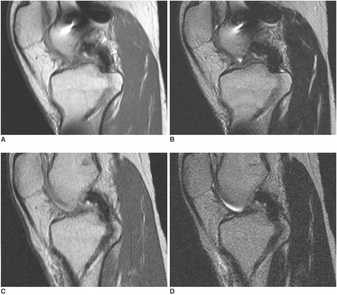
Fig. 1 Proton density-weighted sagittal (A, C) (TR/TE, 2000/20 msec; echo train length, 4) and T2-weighted sagittal (B, D) (TR/TE, 2000/80 msec; echo train length, 4) images of a 21-year-old man who received posterior cruciate ligament reconstruction using autogenous double-loop hamstring tendon (patient #7). Proton density-weighted image obtained 232 days after posterior cruciate ligament reconstruction shows high band-like peripheral signal intensity (arrowheads) in the proximal and middle segments (A). The high signal intensity has disappeared, and the graft shows homogeneous low signal intensity on proton density-weighted image obtained 472 days after posterior cruciat ligament reconstruction (C). The graft shows homogenous low signal intensity on T2-weighted images (B, D).
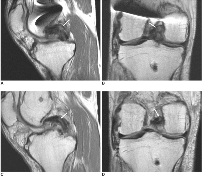
Fig. 2 Proton density-weighted sagittal (A, C) and coronal (B, D) images (TR/TE, 2000/20 msec; echo train length, 4) of a 35-year-old man who received posterior cruciate ligament reconstruction using autogenous double-loop hamstring tendon graft (patient #6). MR images obtained 265 days after posterior cruciate ligament reconstruction (A, B) show high band-like intrasubstance signal intensity (arrows) in the middle segment. The high signal intensity (arrows) persists on MR images obtained 407 days after posterior cruciate ligament reconstruction (C, D). The graft thickness decreases from 15 mm (A) to 12 mm (C).

Fig. 3 Proton density-weighted sagittal (A, C) and coronal (B, D) images (TR/TE, 2000/20 msec; echo train length, 4) of a 36-year-old man who received posterior cruciate ligament reconstruction using Achilles tendon allograft (patient #13). MR images obtained 118 days after posterior cruciate ligament reconstruction (A, B) show high band-like intrasubstance (arrow) and peripheral signal intensity (arrowheads) in the proximal, middle, and distal segments. MR images obtained 773 days after posterior cruciate ligament reconstruction (C, D) show high focal intrasubstance and peripheral signal intensity (arrows) in the proximal and middle segments. The graft thickness increases from 8 mm (A) to 11 mm (C).
Reference
-
1. Höher J, Harner CD, Vogrin TM, Baek GH, Carlin GJ, Woo SL. In situ forces in the posterolateral structures of the knee under posterior tibial loading in the intact and posterior cruciate ligament-deficient knee. J Orthop Res. 1998. 16:675–681.2. Dowd GS. Reconstruction of the posterior cruciate ligament. Indications and results. J Bone Joint Surg. 2004. 86:Br. 480–491.3. Parolie JM, Bergfeld JA. Long-term results of nonoperative treatment of isolated posterior cruciate ligament injuries in the athlete. Am J Sports Med. 1986. 14:35–38.4. Covey CD, Sapega AA. Injuries of the posterior cruciate ligament. J Bone Joint Surg Am. 1993. 75:1376–1386.5. Richter M, Kiefer H, Hehl G, Kinzl L. Primary repair for posterior cruciate ligament injuries. An eight-year followup of fifty-three patients. Am J Sports Med. 1996. 24:298–305.6. Berg EE. Posterior cruciate ligament tibial inlay reconstruction. Arthroscopy. 1995. 11:69–76.7. Miller MD, Bergfeld JA, Fowler PJ, Harner CD, Noyes FR. The posterior cruciate ligament injured knee: principles of evaluation and treatment. Instr Course Lect. 1999. 48:199–207.8. Ahn JH, Chung YS, Oh I. Arthroscopic posterior cruciate ligament reconstruction using the posterior trans-septal portal. Arthroscopy. 2003. 19:101–107.9. Fanelli GC, Giannotti BF, Edson CJ. The posterior cruciate ligament arthroscopic evaluation and treatment. Arthroscopy. 1994. 10:673–688.10. Sanders TG. MR imaging of postoperative ligaments of the knee. Semin Musculoskelet Radiol. 2002. 6:19–33.11. Burns WC 2nd, Draganich LF, Pyevich M, Reider B. The effect of femoral tunnel position and graft tensioning technique on posterior laxity of the posterior cruciate ligament-reconstructed knee. Am J Sports Med. 1995. 23:424–430.12. Munk PL, Vellet AD, Fowler PJ, Miniaci T, Crues JV 3rd. Magnetic resonance imaging of reconstructed knee ligaments. Can Assoc Radiol J. 1992. 43:411–419.13. Irizarry JM, Recht MP. MR imaging of the knee ligaments and the postoperative knee. Radiol Clin North Am. 1997. 35:45–76.14. Sherman PM, Sanders TG, Morrison WB, Schweitzer ME, Leis HT, Nusser CA. MR imaging of the posterior cruciate ligament graft: initial experience in 15 patients with clinical correlation. Radiology. 2001. 221:191–198.15. Mariani PP, Margheritini F, Camillieri G, Bellelli A. Serial magnetic resonance imaging evaluation of the patellar tendon after posterior cruciate ligament reconstruction. Arthroscopy. 2002. 18:38–45.16. Hong SJ, Ahn JM, Ahn JH, Park SW. Postoperative MR findings of the healthy ACL grafts: correlation with second look arthroscopy. Clin Imaging. 2005. 29:55–59.17. Howell SM, Berns GS, Farley TE. Unimpinged and impinged anterior cruciate ligament grafts: MR signal intensity measurements. Radiology. 1991. 179:639–643.18. Cheung Y, Magee TH, Rosenberg ZS, Rose DJ. MRI of anterior cruciate ligament reconstruction. J Comput Assist Tomogr. 1992. 16:134–137.19. Yamato M, Yamagishi T. MRI of patellar tendon anterior cruciate ligament autografts. J Comput Assist Tomogr. 1992. 16:604–607.20. Horton LK, Jacobson JA, Lin J, Hayes CW. MR imaging of anterior cruciate ligament reconstruction graft. AJR Am J Roentgenol. 2000. 175:1091–1097.21. Jansson KA, Karjalainen PT, Harilainen A, Sandelin J, Soila K, Tallroth K, et al. MRI of anterior cruciate ligament repair with patellar and hamstring tendon autografts. Skeletal Radiol. 2001. 30:8–14.22. Canale ST. Campbell's Operative Orthopaedics. 2003. 10th ed. St.Louis: Mosby, Inc;2166.23. Murakami Y, Sumen Y, Ochi M, Fujimoto E, Adachi N, Ikuta Y. MR evaluation of human anterior cruciate ligament autograft on oblique axial imaging. J Comput Assist Tomogr. 1998. 22:270–275.24. Trattnig S, Rand T, Czerny C, Stocker R, Breitenseher M, Kainberger F, et al. Magnetic resonance imaging of the postoperative knee. Top Magn Reson Imaging. 1999. 10:221–236.25. Buess E, Imhoff AB, Hodler J. Knee evaluation in two systems and magnetic resonance imaging after operative treatment of posterior cruciate ligament injuries. Arch Orthop Trauma Surg. 1996. 115:307–312.
- Full Text Links
-
- Actions
-
Cited
- CITED
-
- Close
- Share
-
- Similar articles
-
- Ganglion Cyst of the Posterior Cruciate Ligament: A Case Report
- Reconstruction of the Posterior Cruciate Ligament Using the Medial Meniscus
- Arthroscopic Reduction and Internal Fixation of Bony Avulsion of the Posterior Cruciate Ligament
- Symptomatic Calcific Deposition in Posterior Cruciate Ligament of the Knee
- Ganglion of the Posterior Cruciate Ligament: 1 case report
