Calcifying Aponeurotic Fibroma with Osseous Involvement of the Finger: a Case Report with Radiologic and US Findings
- Affiliations
-
- 1Department of Radiology, Asan Foundation, GangNeung Asan Hospital, University of Ulsan College of Medicine, Gangneung, Korea. sjchoi@gnah.co.kr
- 2Department of Pathology, Asan Foundation, GangNeung Asan Hospital, University of Ulsan College of Medicine, Gangneung, Korea.
- KMID: 1734282
- DOI: http://doi.org/10.3348/kjr.2008.9.1.91
Abstract
- Calcifying aponeurotic fibroma is a rare soft tissue tumor that occurs in the distal extremities of children and adolescents. We report ultrasound and X-ray findings of a calcifying aponeurotic fibroma in the finger of a 36-year-old woman, associated with distal phalangeal bone involvement.
MeSH Terms
Figure
-
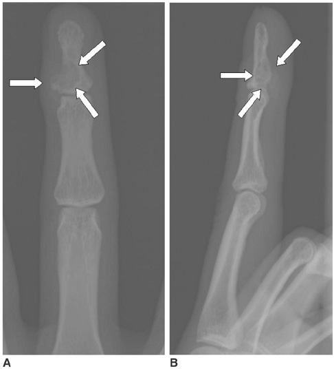
Fig. 1 Left middle finger AP (A) and lateral (B) views show eccentrically located well-defined osteolytic lesion in the base of the distal phalanx (arrows). Calcific foci are noted in the mass and soft tissue mass component is obvious. On these radiographs, soft tissue mass with large cortical erosion is indistinguishable with eccentrically locating osteolytic mass with soft tissue extension.
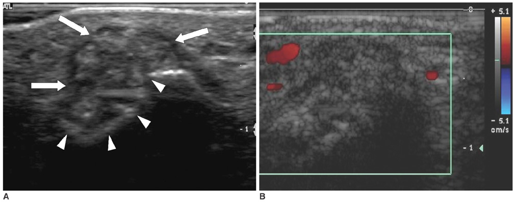
Fig. 2 A. High-resolution US demonstrates lobulated soft tissue mass (arrows) with internal punctate hyper-echoic foci (calcifications). The mass is adhered and scalloped the cortex of the phalanx (arrowheads). B. Color Doppler US shows hypo-vascularity of the mass.

Fig. 3 Histological section shows scattered calcifications (solid arrows) with surrounding chondroid differentiation (arrowheads) on the background of the fibrosis with interlacing bundles of spindle cells (open arrows) (Hematoxylin and Eosin staining, ×40).
Reference
-
1. Karasick D, O'Hara AE. Juvenile aponeurotic fibroma. A review and report of a case with osseous involvement. Radiology. 1977. 123:725–726.2. Robbin MR, Murphey MD, Temple HT, Kransdorf MJ, Choi JJ. Imaging of musculoskeletal fibromatosis. Radiographics. 2001. 21:585–600.3. Rahmi M, Chakkouri K, Cohen D, Hassoun J, Trafeh M. Juvenile aponeurotic fibroma. A case report with a review of the literature. Chir Main. 2002. 21:33–35.4. DeSimone RS, Zielinski CJ. Calcifying aponeurotic fibroma of the hand. J Bone Joint Surg Am. 2001. 83:586–588.5. Parker WL, Beckenbaugh RR, Amrami KK. Calcifying aponeurotic fibroma of the hand: radiologic differentiation from giant cell tumors of the tendon sheath. J Hand Surg (Am). 2006. 31:1024–1028.6. Goldman RL. The cartilage analogue of fibromatosis (aponeurotic fibroma). Further observations based on 7 new cases. Cancer. 1970. 26:1325–1331.7. Kwak HS, Lee SY, Kim JR, Lee KB. MR imaging of calcifying aponeurotic fibroma of the thigh. Pediatr Radiol. 2004. 34:438–440.8. Carroll RE. Juvenile aponeurotic fibroma. Hand Clin. 1987. 3:219–224.
