Korean J Radiol.
2014 Aug;15(4):538-539. 10.3348/kjr.2014.15.4.538.
RE: Hemangiopericytoma of the Greater Omentum: A Potential Imaging Pitfall and Cause of Repeatedly Unsuccessful Angiographic Embolization
- Affiliations
-
- 1Medical and Surgical Sciences and Translational Medicine, Faculty of Medicine and Psychology "Sapienza", St. Andrea Hospital, Rome 00189, Italy. aresedo1992@yahoo.it
- KMID: 1731060
- DOI: http://doi.org/10.3348/kjr.2014.15.4.538
Abstract
- No abstract available.
MeSH Terms
Figure
-

Fig. 1 Contrast-enhanced CT scan of abdomen showed aneurysm of 1.7 × 2 cm arising from short gastric artery (red arrow).
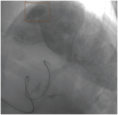
Fig. 2 Superselective splenic arteriography confirmed aneurysmatic nature of lesion (orange square).
Reference
-
1. Kulkarni CB, Moorthy S, Pullara SK, Kannan RR. Endovascular treatment of aneurysm of splenic artery arising from splenomesentric trunk using stent graft. Korean J Radiol. 2013; 14:931–934.2. Goldberger RE, Schein CJ. Hemangiopericytoma of the omentum. Report of a case with a unique presentation and review of the literature. Am Surg. 1968; 34:291–229.3. Morris-Stiff G, Falk GA, Joyce D, Rubin B, Chalikonda S. Primary omental haemangiopericytoma. BMJ Case Rep. 2011; doi: 10.1136/bcr.03.2011.4041.4. Crusco F, Chiodi M, Pugliese F, Mosca S, Fischer MJ, Lupattelli L. Benign omental hemangiopericytoma presenting with hemoperitoneum: radiologic findings. AJR Am J Roentgenol. 2005; 184:3 Suppl. S67–S69.
- Full Text Links
-
- Actions
-
Cited
- CITED
-
- Close
- Share
-
- Similar articles
-
- Hemangiopericytoma of the Greater Omentum Mimicking Ovarian Tumor: a Case Report
- A Rare Metastatic Liver Cancer: Hemangiopericytoma
- Spinal Hemangiopericytoma Which Needed Intraoperative Embolization due to Unexpected Bleeding
- Diffuse Hemangiomatosis in the Intra-Abdominal Cavity Mimicking Peritoneal Metastasis: A Case Report
- CT Findings of Primary Torsion of the Greater Omentum with Segmental Infarction: Case Report
