Korean J Gastroenterol.
2014 Mar;63(3):187-190. 10.4166/kjg.2014.63.3.187.
A Case of Primary Retroperitoneal Mucinous Cystadenoma Arising from the Retropancreatic Area
- Affiliations
-
- 1Division of Gastroenterology and Hepatology, Department of Internal Medicine, Yeungnam University College of Medicine, Daegu, Korea. tnkim@yu.ac.kr
- KMID: 1730931
- DOI: http://doi.org/10.4166/kjg.2014.63.3.187
Abstract
- Primary retroperitoneal mucinous cystadenoma is an extremely uncommon tumor, even though mucinous cystadenoma often develops in the ovary and less frequently in the pancreas. A 21-year-old female was admitted to our hospital due to severe abdominal pain. A well-demarcated, oval shaped cystic tumor at the retropancreatic area with displacement of the pancreas and surrounding major vessels was observed on CT and MRI. Exploratory laparotomy was performed, and complete excision of the entire cyst was performed without complication. The pathologic finding was consistent with primary retropancreatic mucinous cystadenoma. To the best of our knowledge, this report is the first to describe a case of retropancreatic mucinous cystadenoma arising from the retropancreatic area in Korea.
Keyword
MeSH Terms
-
Antibodies/metabolism
Cystadenoma, Mucinous/*diagnosis/pathology/surgery
Female
Humans
Magnetic Resonance Imaging
Mucin 5AC/immunology
Mucin-2/immunology
Ovarian Neoplasms/*diagnosis/pathology/surgery
Retroperitoneal Neoplasms/*diagnosis/pathology/surgery
Tomography, X-Ray Computed
Young Adult
Antibodies
Mucin 5AC
Mucin-2
Figure
-
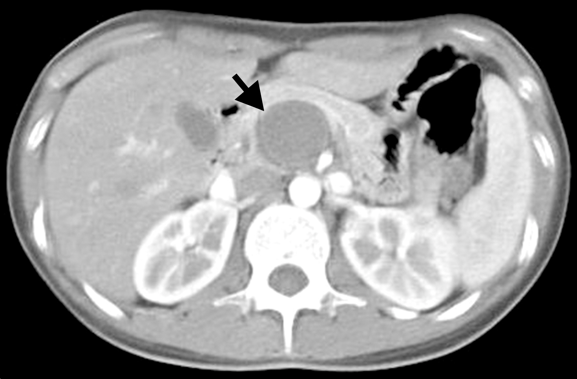
Fig. 1. CT scan of the abdomen. A well-demarcated oval shaped cystic tumor measuring 3.0×6.0 cm in size at the retropancreatic area with displacement of the pancreas and surrounding major vessels was observed (black arrow).

Fig. 2. MRI of the abdomen. The cyst consisted of protein rich liquid contents with irregular thickening of the cyst wall (white arrows).
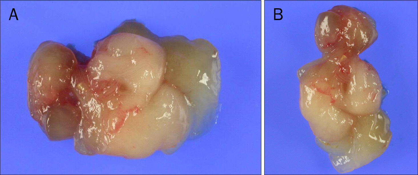
Fig. 3. Gross finding of the resected specimen. The cystic mass measured 5.5 × 3.5 cm in size and weighed 11.8 g, and consisted of thick mucus material on the cut surface.

Fig. 4. Microscopic finding. (A) The cyst wall was lined with a single layer of mucinous epithelial cells (H&E, ×200). (B) On immunohistochemical staining, MUC2 and MUC5AC antibodies were positive (×200).
Reference
-
References
1. Metaxas G, Tangalos A, Pappa P, Papageorgiou I. Mucinous cystic neoplasms of the mesentery: a case report and review of the literature. World J Surg Oncol. 2009; 7:47.
Article2. Cho HY, Kim YH, Kim JW, Choi SJ, Song TB. Primary retroperitoneal mucinous cystadenoma mimicking a left ovarian tumor in pregnant woman: a case report. J Womens Med. 2010; 3:170–173.
Article3. Lai KKT, Chan YYR, Chin ACW, et al. Primary retroperitoneal mucinous cystadenoma in a 52-year-old man. J HK Coll Radiol. 2004; 7:223–225.4. Kim GY, Choi DH, Lim YC, et al. Retroperitoneal mucinous cystadenoma. J Korean Surg Soc. 2008; 74:79–82.5. Subramony C, Habibpour S, Hashimoto LA. Retroperitoneal mucinous cystadenoma. Arch Pathol Lab Med. 2001; 125:691–694.
Article6. Arribas D, Cay A, Latorre A, Córdoba E, Martínez F, Lagos J. Retroperitoneal mucinous cystadenoma. Arch Gynecol Obstet. 2004; 270:292–293.
Article7. Tapper EB, Shrewsberry AB, Oprea G, Majmudar B. A unique benign mucinous cystadenoma of the retroperitoneum: a case report and review of the literature. Arch Gynecol Obstet. 2010; 281:167–169.
Article8. Yang DM, Jung DH, Kim H, et al. Retroperitoneal cystic masses: CT, clinical, and pathologic findings and literature review. Radiographics. 2004; 24:1353–1365.
Article9. Shiau JP, Wu CT, Chin CC, Chuang CK. Longterm survival after hand-assisted laparoscopic approach of primary retroperitoneal mucinous cystadenocarcinoma in male: case report and review of literature. Eur Surg. 2013; 45:106–109.
Article10. Yan SL, Lin H, Kuo CL, Wu HS, Huang MH, Lee YT. Primary retroperitoneal mucinous cystadenoma: report of a case and review of the literature. World J Gastroenterol. 2008; 14:5769–5772.
Article
