En-masse retraction with a preformed nickel-titanium and stainless steel archwire assembly and temporary skeletal anchorage devices without posterior bonding
- Affiliations
-
- 1Department of Orthodontics, Postgraduate School of Dentistry, The Catholic University of Korea, Seoul, Korea.
- 2Department of Orthodontics, School of Dentistry, Kyung Hee University, Seoul, Korea. bravortho@gmail.com
- 3Department of Orthodontics, Ajou University School of Medicine, Suwon, Korea.
- 4Division of Orthodontics, Department of Orofacial Science, University of California, San Francisco, CA, USA.
- KMID: 1726417
- DOI: http://doi.org/10.4041/kjod.2014.44.5.236
Abstract
OBJECTIVE
To evaluate the therapeutic effects of a preformed assembly of nickel-titanium (NiTi) and stainless steel (SS) archwires (preformed C-wire) combined with temporary skeletal anchorage devices (TSADs) as the sole source of anchorage and to compare these effects with those of a SS version of C-wire (conventional C-wire) for en-masse retraction.
METHODS
Thirty-one adult female patients with skeletal Class I or II dentoalveolar protrusion, mild-to-moderate anterior crowding (3.0-6.0 mm), and stable Class I posterior occlusion were divided into conventional (n = 15) and preformed (n = 16) C-wire groups. All subjects underwent first premolar extractions and en-masse retraction with pre-adjusted edgewise anterior brackets, the assigned C-wire, and maxillary C-tubes or C-implants; bonded mesh-tube appliances were used in the mandibular dentition. Differences in pretreatment and post-retraction measurements of skeletal, dental, and soft-tissue cephalometric variables were statistically analyzed.
RESULTS
Both groups showed full retraction of the maxillary anterior teeth by controlled tipping and space closure without altered posterior occlusion. However, the preformed C-wire group had a shorter retraction period (by 3.2 months). Furthermore, the maxillary molars in this group showed no significant mesialization, mesial tipping, or extrusion; some mesialization and mesial tipping occurred in the conventional C-wire group.
CONCLUSIONS
Preformed C-wires combined with maxillary TSADs enable simultaneous leveling and space closure from the beginning of the treatment without maxillary posterior bonding. This allows for faster treatment of dentoalveolar protrusion without unwanted side effects, when compared with conventional C-wire, evidencing its clinical expediency.
MeSH Terms
Figure
-
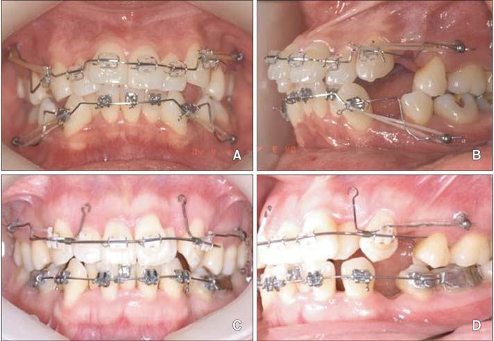
Figure 1 Temporary skeletal anchorage device-dependent en-masse retraction mechanics. A and B, Biocreative therapy with conventional C-wires; note the bends and steps in the archwire. C and D, Biocreative therapy with preformed C-wires.

Figure 2 Temporary skeletal anchorage devices used in this study. A and B, Two-component C-implants. C and D, C-tubes.
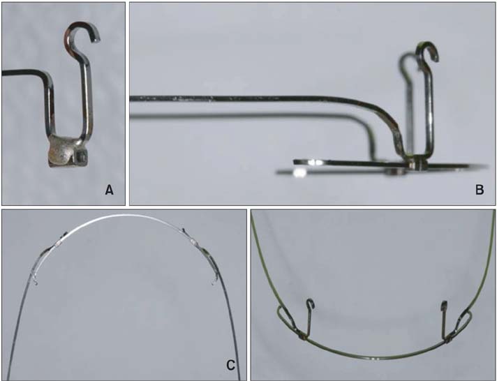
Figure 3 Preformed C-wire fabrication. A, Bending of stainless steel (SS) archwire for the retraction hooks and soldering of the crimpable stop tubes. B, Insertion of nickel-titanium archwire into the tubes and contouring of the hooks for passive sliding according to the height of the C-tubes. C, Contouring of the remaining SS section. D, Final configuration of preformed C-wire.
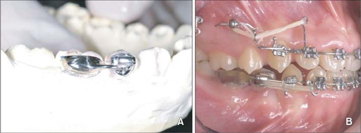
Figure 4 (A) Bonded mesh-tube appliance (BMTA) on the mandibular arch. The BMTA is comprised of a 0.022-inch single tube on the mandibular second premolar connected to metal mesh on the mandibular first molar (B).
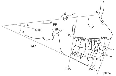
Figure 5 Soft tissue and skeletal cephalometric measurements. 1, Upper lip to E-line (UL to E-line); 2, lower lip to E-line (LL to E-line); 3, sella-nasion to palatal plane angle (SN-PP); 4, SN to anatomic occlusal plane angle (SN-Occ); 5, SN to mandibular plane angle (SN-MP); 6, pterygoid vertical plane to A point distance (PTV-A); 7, pterygoid vertical plane to B point distance (PTV-B); 8, lower anterior facial height (anterior nasal spine to menton; ANS-Me).
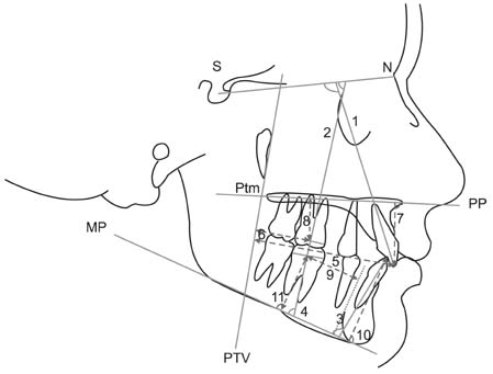
Figure 6 Dental cephalometric measurements. 1, Sellanasion to maxillary incisor angle (SN-U1); 2, SN to maxillary first molar angle (SN-U6); 3, mandibular plane to mandibular incisor angle (MP-L1); 4, mandibular plane to mandibular first molar angle (MP-L6); 5, pterygoid vertical plane to maxillary incisor tip distance (PTV-U1); 6, pterygoid vertical plane to maxillary first molar centroid distance (PTV-U6); 7, palatal plane to maxillary incisor tip distance (PP-U1); 8, palatal plane to maxillary first molar centroid distance (PP-U6); 9, mandibular lingual cortex to mandibular first molar centroid distance (LC-L6); 10, mandibular plane to mandibular incisor tip distance (MP-L1v); 11, mandibular plane to mandibular first molar centroid distance (MP-L6v).
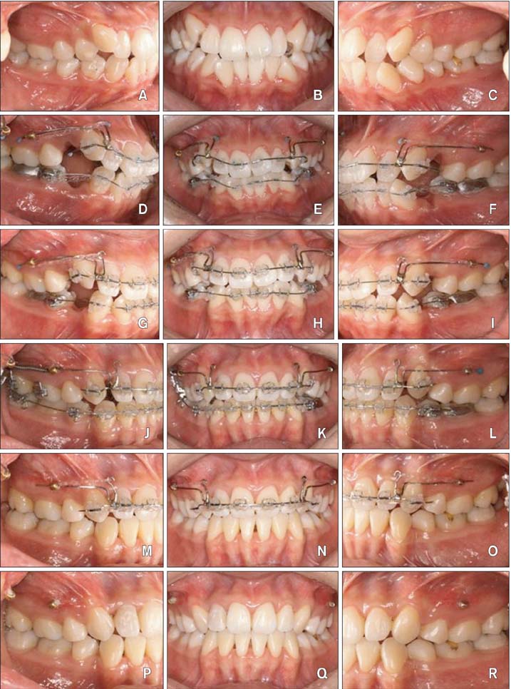
Figure 7 Therapeutic progress with one preformed C-wire. A to C, Pretreatment. D to F, Immediately after bonding. G to I, Three months after treatment. J to L, Nine months after treatment. M to O, Fifteen months after treatment. P to R, Posttreatment (nineteen month after treatment).
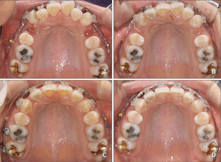
Figure 8 Occlusal views showing relief of crowding and retraction of the maxillary anterior teeth. A, Immediately after bonding. B, Three months after bonding. C, Nine months after bonding. D, Fifteen months after bonding.
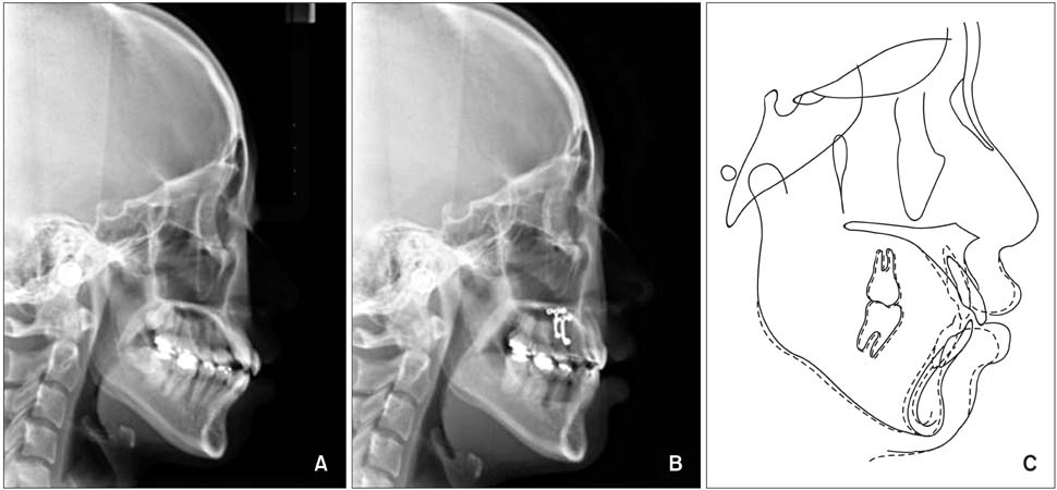
Figure 9 Lateral cephalograms and superimposed tracings. A, Pretreatment lateral cephalogram. B, Post-retraction lateral cephalogram. C, Superimposition of the pretreatment (solid line) and post-retraction (dotted line) tracings.
Cited by 3 articles
-
Displacement pattern of the anterior segment using antero-posterior lingual retractor combined with a palatal plate
Kyung-Won Seo, Soon-Yong Kwon, Kyung A Kim, Ki-Ho Park, Seong-Hun Kim, Hyo-Won Ahn, Gerald Nelson
Korean J Orthod. 2015;45(6):289-298. doi: 10.4041/kjod.2015.45.6.289.Type of tooth movement during en masse retraction of the maxillary anterior teeth using labial versus lingual biocreative therapy in adults: A randomized clinical trial
Mais M. Sadek, Noha E. Sabet, Islam T. Hassan
Korean J Orthod. 2019;49(6):381-392. doi: 10.4041/kjod.2019.49.6.381.A new rationale for preservation of the mandibular third molar in orthognathic patients with missing molars
Un-Bong Baik, Yoon-Ji Kim, Hwa-Sung Chae, Je-Uk Park, Stefania Julian, Junji Sugawara, Ui-Lyong Lee
J Korean Assoc Oral Maxillofac Surg. 2022;48(1):63-67. doi: 10.5125/jkaoms.2022.48.1.63.
Reference
-
1. Bennett JC, McLaughlin RP. Controlled space closure with a preadjusted appliance system. J Clin Orthod. 1990; 24:251–260.2. Klontz HA. Tweed-Merrifield sequential directional force treatment. Semin Orthod. 1996; 2:254–267.3. Kanomi R. Mini-implant for orthodontic anchorage. J Clin Orthod. 1997; 31:763–767.4. Yao CC, Lai EH, Chang JZ, Chen I, Chen YJ. Comparison of treatment outcomes between skeletal anchorage and extraoral anchorage in adults with maxillary dentoalveolar protrusion. Am J Orthod Dentofacial Orthop. 2008; 134:615–624.
Article5. Block MS, Hoffman DR. A new device for absolute anchorage for orthodontics. Am J Orthod Dentofacial Orthop. 1995; 107:251–258.
Article6. Chung KR, Kim YS, Linton JL, Lee YJ. The miniplate with tube for skeletal anchorage. J Clin Orthod. 2002; 36:407–412.7. Ahn HW, Chung KR, Kang SM, Lin L, Nelson G, Kim SH. Correction of dental Class III with posterior open bite by simple biomechanics using an anterior C-tube miniplate. Korean J Orthod. 2012; 42:270–278.
Article8. Chung KR, Cho JH, Kim SH, Kook YA, Cozzani M. Unusual extraction treatment in Class II division 1 using C-orthodontic mini-implants. Angle Orthod. 2007; 77:155–166.
Article9. Chung KR, Choo H, Lee JH, Kim SH. Atypical orthodontic extraction pattern managed by differential en-masse retraction against a temporary skeletal anchorage device in the treatment of bimaxillary protrusion. Am J Orthod Dentofacial Orthop. 2011; 140:423–432.
Article10. Chung KR, Jeong DM, Kim SH, Ko YI, Nelson G. En-masse retraction dependent on a temporary skeletal anchorage device without posterior bonding or banding in an adult with severe bidentoalveolar protrusion: seven years posttreatment. Am J Orthod Dentofacial Orthop. 2012; 141:484–494.
Article11. Kim SH, Hwang YS, Ferreira A, Chung KR. Analysis of temporary skeletal anchorage devices used for en-masse retraction: a preliminary study. Am J Orthod Dentofacial Orthop. 2009; 136:268–276.
Article12. Chung KR, Kim SH, Kook YA. C-orthodontic mini-implant. In : Cope JB, editor. OrthoTADs book: Clinical guideline and atlas. Dallas, TX: Under Dog Media;2007. p. 248.13. Chung KR, Kim SH, Kook YA. The C-orthodontic micro-implant. J Clin Orthod. 2004; 38:478–486.14. Giancotti A, Greco M. Modified sliding mechanics in extraction cases with a bidimensional approach. Prog Orthod. 2010; 11:157–165.
Article15. Mandall N, Lowe C, Worthington H, Sandler J, Derwent S, Abdi-Oskouei M, et al. Which orthodontic archwire sequence? A randomized clinical trial. Eur J Orthod. 2006; 28:561–566.
Article16. Garrec P, Tavernier B, Jordan L. Evolution of flexural rigidity according to the cross-sectional dimension of a superelastic nickel titanium orthodontic wire. Eur J Orthod. 2005; 27:402–407.
Article17. Gurgel JA, Kerr S, Powers JM, LeCrone V. Force-deflection properties of superelastic nickel-titanium archwires. Am J Orthod Dentofacial Orthop. 2001; 120:378–382.
Article18. Sia S, Shibazaki T, Koga Y, Yoshida N. Experimental determination of optimal force system required for control of anterior tooth movement in sliding mechanics. Am J Orthod Dentofacial Orthop. 2009; 135:36–41.
Article19. Mo SS, Kim SH, Sung SJ, Chung KR, Chun YS, Kook YA, et al. Factors controlling anterior torque during C-implant-dependent en-masse retraction without posterior appliances. Am J Orthod Dentofacial Orthop. 2011; 140:72–80.
Article20. Mo SS, Kim SH, Sung SJ, Chung KR, Chun YS, Kook YA, et al. Factors controlling anterior torque with C-implants depend on en-masse retraction without posterior appliances: biocreative therapy type II technique. Am J Orthod Dentofacial Orthop. 2011; 139:e183–e191.
- Full Text Links
-
- Actions
-
Cited
- CITED
-
- Close
- Share
-
- Similar articles
-
- Finite element analysis of the effects of different archwire forms and power arm positions on maxillary incisors in en masse retraction using fixed lingual orthodontic appliances
- The skeletal cortical anchorage using titanium microscrew implants
- Effectiveness of anchorage with temporary anchorage devices during anterior maxillary tooth retraction: A randomized clinical trial
- Mechanical properties of nickel titanium and steel alloys under stress- strain test
- Effect of archwire stiffness and friction on maxillary posterior segment displacement during anterior segment retraction: A three-dimensional finite element analysis
