A Case of Mantle Cell Lymphoma with Meningioma
- Affiliations
-
- 1Department of Internal Medicine, Chungnam National University School of Medicine, Daejeon, Korea. mhs1357@cnuh.co.kr
- KMID: 1718468
- DOI: http://doi.org/10.4166/kjg.2012.60.1.56
Abstract
- Mantle cell lymphoma (MCL) is an uncommon type of gastrointestinal lymphoma. MCL is a distinct subtype of B-cell non-Hodgkin lymphomas. The major subtype of MCL is characterized by the presence of multiple lymphomatous polyposis (MLP), in which multiple polyps are observed along the gastrointestinal tract. The malignant cells express pan B-cell marker and the T-cell marker cluster of differentiation 5. The chromosomal translocation t(11;14)(q13;q32) that causes cyclin D1 overexpression is commonly observed on the cytogenetic analysis of MCL. Survival improvement has recently been achieved for patient with MCL by the successful introduction of monoclonal antibodies and dose-intensified approaches for treatment, including autologous stem cell transplantation strategies. Some reports suggest that there is an increased incidence of second malignancies in patients with MCL or lymphoma. We report a case of MCL involving the colon; the patient was a 60-year-old man who complained of low abdominal discomfort during defecation. During the workup, a meningioma was unexpectedly discovered. On analysis, the tumor was found to be a t(11;14)-negative and non-MLP-type MCL.
MeSH Terms
-
Chromosomes, Human, Pair 11
Chromosomes, Human, Pair 14
Cyclin D1/metabolism
Humans
Lymphoma, Mantle-Cell/*diagnosis/genetics/metabolism
Magnetic Resonance Imaging
Male
Meningeal Neoplasms/complications/*diagnosis/pathology
Meningioma/complications/*diagnosis/pathology
Middle Aged
Positron-Emission Tomography
Translocation, Genetic
Figure
-
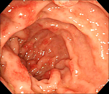
Fig. 1 Endoscopic findings. Inflammatory mucosal changes were noted in the entire large intestine, especially in the rectum. Polypectomy was performed in the rectal region.
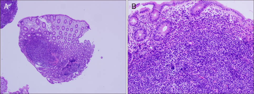
Fig. 2 Microscopic findings (H&E). Neoplastic cells had infiltrated the lamina propria of the colon (A, ×40). Intraepithelial B lymphocytes were forming the lymphoepithelium. The tumor cells were small-to-medium sized (B, ×200).
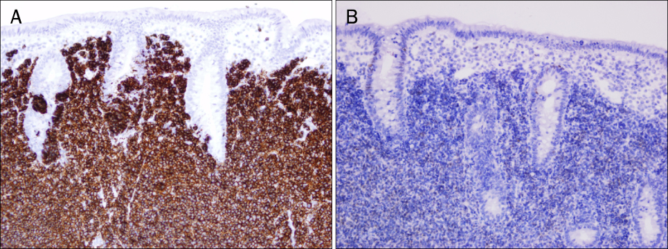
Fig. 3 Immunohistochemical staining. Tumore cells were strongly positive for CD20 and exhibit CD5 coexpression (A, ×200). Weak staining was observed for cyclin D1 in the nuclei of the tumor cells (B, ×200).
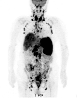
Fig. 4 PET image showed multiple hypermetabolic lesions in lymph nodes in the entire body, splenomegaly, and colon of terminal ileum, cecum, and rectum.
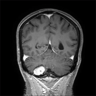
Fig. 5 T2-weighted brain MRI scan showed an approximately 2.6×2.5×1.7 cm meningioma in the right cerebellar convexity.
Reference
-
1. Loehr WJ, Mujahed Z, Zahn FD, Gray GF, Thorbjarnarson B. Primary lymphoma of the gastrointestinal tract: a review of 100 cases. Ann Surg. 1969. 170:232–238.2. Kim DH, Sohn SK, Jung JT, et al. Mantle cell lymphoma presented as multiple lymphomatous polyposis: clinical review of 4 cases. Korean J Med. 2000. 59:413–422.3. Lim HJ, Choi P, Hur JW, et al. A case of mantle cell lymphoma presenting as multiple lymphomatous polyposis involving skeletal muscles. Korean J Gastrointest Endosc. 2003. 27:15–20.4. Hemminki K, Lenner P, Sundquist J, Bermejo JL. Risk of subsequent solid tumors after non-Hodgkin's lymphoma: effect of diagnostic age and time since diagnosis. J Clin Oncol. 2008. 26:1850–1857.5. Barista I, Cabanillas F, Romaguera JE, et al. Is there an increased rate of additional malignancies in patients with mantle cell lymphoma? Ann Oncol. 2002. 13:318–322.6. Lolli E, Matteoni R, Barbieri A, D'Ambrosi M, Archontakis F. Non-Hodgkin's lymphoma, renal carcinoma, and meningioma. A clinical case. Ann Ital Chir. 1998. 69:101–104.7. Dreyling M, Hiddemann W. European MCL Network. Current treatment standards and emerging strategies in mantle cell lymphoma. Hematology Am Soc Hematol Educ Program. 2009. 542–551.8. Isaacson PG, MacLennan KA, Subbuswamy SG. Multiple lymphomatous polyposis of the gastrointestinal tract. Histopathology. 1984. 8:641–656.9. Harris NL, Jaffe ES, Stein H, et al. A revised European-American classification of lymphoid neoplasms: a proposal from the International Lymphoma Study Group. Blood. 1994. 84:1361–1392.10. Tagle P, Villanueva P, Torrealba G, Huete I. Intracranial metastasis or meningioma? An uncommon clinical diagnostic dilemma. Surg Neurol. 2002. 58:241–245.11. Norton AJ, Matthews J, Pappa V, et al. Mantle cell lymphoma: natural history defined in a serially biopsied population over a 20-year period. Ann Oncol. 1995. 6:249–256.12. Ruskoné-Fourmestraux A, Delmer A, Lavergne A, et al. Groupe D'étude des Lymphomes Digestifs. Multiple lymphomatous polyposis of the gastrointestinal tract: prospective clinicopathologic study of 31 cases. Gastroenterology. 1997. 112:7–16.13. Zucca E, Stein H, Coiffier B. European Lymphoma Task Force (ELTF): Report of the workshop on mantle cell lymphoma (MCL). Ann Oncol. 1994. 5:507–511.14. Swerdlow SH, Williams ME. From centrocytic to mantle cell lymphoma: a clinicopathologic and molecular review of 3 decades. Hum Pathol. 2002. 33:7–20.15. Hoster E, Dreyling M, Klapper W, et al. German Low Grade Lymphoma Study Group (GLSG). European Mantle Cell Lymphoma Network. A new prognostic index (MIPI) for patients with advanced-stage mantle cell lymphoma. Blood. 2008. 111:558–565.16. Leonard JP, Schattner EJ, Coleman M. Biology and management of mantle cell lymphoma. Curr Opin Oncol. 2001. 13:342–347.17. Herrmann A, Hoster E, Zwingers T, et al. Improvement of overall survival in advanced stage mantle cell lymphoma. J Clin Oncol. 2009. 27:511–518.
- Full Text Links
-
- Actions
-
Cited
- CITED
-
- Close
- Share
-
- Similar articles
-
- A Case of Primary Mantle Cell Lymphoma on the Conjunctiva
- A Case of Primary Cutaneous Marginal Zone B-cell Lymphoma
- A Case of Mantle Cell Lymphoma Treated with Autologous Stem Cell Transplantation and Rituximab
- Mantle cell lymphoma: a model for risk-adapted treatment approach
- Circulating Lymphoma Cells in the Peripheral Blood from 4 Cases of Mantle and T Cell Types of Non-Hodgkin's Lymphoma: Light and Electron Microscopic Morphology
