Rapid Intra-Hepatic Dissemination of Hepatocellular Carcinoma with Pulmonary Metastases Following Combined Loco-Regional Therapy
- Affiliations
-
- 1Department of Diagnostic Radiology, Tan Tock Seng Hospital, Singapore. druei@yahoo.com
- 2Yong Loo Lin School of Medicine, National University of Singapore, Singapore.
- KMID: 1715769
- DOI: http://doi.org/10.3348/kjr.2013.14.4.640
Abstract
- This manuscript describes an unusual case of rapid intra-hepatic dissemination of hepatocellular carcinoma with pulmonary metastases occurring 1 month after combined chemoembolization and radiofrequency ablation. Inferior vena cava and portal vein invasion tumor thrombus was also detected, possibly accounting for the mechanism of disease dissemination route of disease.
MeSH Terms
-
Aged, 80 and over
Antineoplastic Agents/administration & dosage/*adverse effects
Biopsy
Carcinoma, Hepatocellular/diagnosis/*secondary/therapy
Catheter Ablation/*adverse effects
Chemoembolization, Therapeutic/*adverse effects
Cone-Beam Computed Tomography
Fatal Outcome
Humans
Liver Neoplasms/*pathology/therapy
Lung Neoplasms/diagnosis/*secondary
Male
Antineoplastic Agents
Figure
-
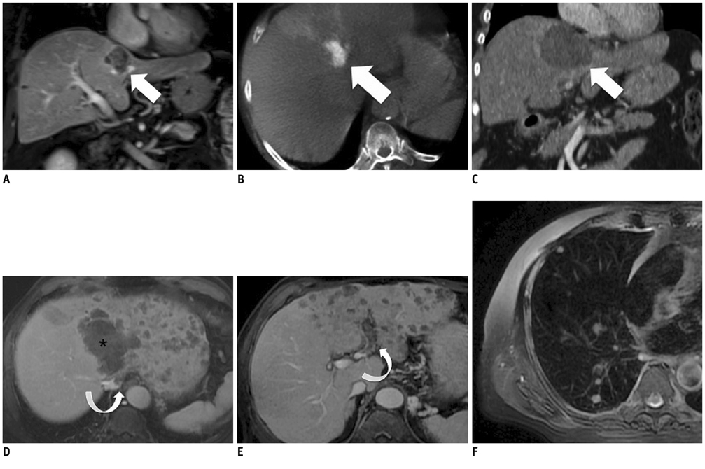
Fig. 1 Multimodality images (A) before TACE, (B) during TACE, (C-E) 4 weeks after TACE. A. Contrast enhanced coronal T1 weighted MR image of solitary segment IV tumor (white arrow) showing areas of heterogeneous central enhancement. No other tumors were present in liver. B. Axial cone-beam CT image after segmental TACE with DC beads infused via middle hepatic artery showing dense tumoral uptake of beads (white arrow) with background staining and beads deposition in segment IV parenchyma. C. Contrast enhanced CT of liver reconstructed in coronal plane, obtained immediately post-RFA, showing ablation zone (white arrow) to be encompassing tumor (compare with A) with at least 1 cm margin. No other tumors in rest of liver and lung bases were seen. D, E. Contrast enhanced T1 weighted axial images MR images, obtained 4 weeks after RFA. D. Multiple small rim enhancing nodules of similar sizes were present in left lobe and around ablation zone (*), with sparing of right lobe. Small tumor thrombus in inferior vena cava was present (curved white arrow). E. Tumor thrombus in left portal vein (curve arrow) was present. Similar findings of multiple small nodules of similar sizes scattered in left lobe with sparing of right lobe were also noted. F. Ax T2 image of right lung obtained 4 weeks after RFA showed several new pulmonary nodules, consistent with metastases. Left lung base nodules of similar sizes were also present (not shown). These nodules were not present on CT performed immediate post-RFA. TACE = transarterial chemo-embolization, RFA = radiofrequency ablation
Reference
-
1. Nicoli N, Casaril A, Abu Hilal M, Mangiante G, Marchiori L, Ciola M, et al. A case of rapid intrahepatic dissemination of hepatocellular carcinoma after radiofrequency thermal ablation. Am J Surg. 2004; 188:165–167.2. Ruzzenente A, Manzoni GD, Molfetta M, Pachera S, Genco B, Donataccio M, et al. Rapid progression of hepatocellular carcinoma after Radiofrequency Ablation. World J Gastroenterol. 2004; 10:1137–1140.3. Mori Y, Tamai H, Shingaki N, Moribata K, Shiraki T, Deguchi H, et al. Diffuse intrahepatic recurrence after percutaneous radiofrequency ablation for solitary and small hepatocellular carcinoma. Hepatol Int. 2009; 3:509–515.4. Kotoh K, Enjoji M, Arimura E, Morizono S, Kohjima M, Sakai H, et al. Scattered and rapid intrahepatic recurrences after radio frequency ablation for hepatocellular carcinoma. World J Gastroenterol. 2005; 11:6828–6832.
- Full Text Links
-
- Actions
-
Cited
- CITED
-
- Close
- Share
-
- Similar articles
-
- Multiple Bone Metastases in a Patient with Small Hepatocellular Carcinoma
- Curative Loco-regional Therapies for Early Hepatocellular Carcinoma: Is Combination Effective?
- High-Dose Vitamin C Promotes Regression of Multiple Pulmonary Metastases Originating from Hepatocellular Carcinoma
- The position of loco-regional therapy in the management of hepatocellular carcinoma with extrahepatic metastases
- Can metronomic chemotherapy be an alternative to sorafenib in advanced hepatocellular carcinoma?
