Pre-Treatment Diffusion-Weighted MR Imaging for Predicting Tumor Recurrence in Uterine Cervical Cancer Treated with Concurrent Chemoradiation: Value of Histogram Analysis of Apparent Diffusion Coefficients
- Affiliations
-
- 1Department of Radiology, Chonnam National University Hwasun Hospital, Chonnam National University Medical School, Hwasun 519-763, Korea.
- 2Department of Radiology, Chonnam National University Hospital, Chonnam National University Medical School, Gwangju 501-757, Korea. kjradsss@dreamwiz.com
- 3Center for Aging and Geriatrics, Chonnam National University Hospital, Chonnam National University Medical School, Gwangju 501-757, Korea.
- 4Department of Obstetrics and Gynecology, Chonnam National University Hwasun Hospital, Chonnam National University Medical School, Hwasun 519-763, Korea.
- KMID: 1715766
- DOI: http://doi.org/10.3348/kjr.2013.14.4.616
Abstract
OBJECTIVE
To evaluate the value of apparent diffusion coefficient (ADC) histogram analysis for predicting tumor recurrence in patients with uterine cervical cancer treated with chemoradiation therapy (CRT).
MATERIALS AND METHODS
Our institutional review board approved this retrospective study and waived informed consent from each patient. Forty-two patients (mean age, 56 +/- 14 years) with biopsy-proven uterine cervical squamous cell carcinoma who underwent both pre-treatment pelvic magnetic resonance imaging with a 3.0 T magnetic resonance scanner and concurrent CRT were included. All patients were followed-up for more than 6 months (mean, 36.4 +/- 11.9 months; range 9.0-52.8 months) after completion of CRT. Baseline ADC parameters (mean ADC, 25th percentile, 50th percentile, and 75th percentile ADC values) of tumors were calculated and compared between the recurrence and no recurrence groups.
RESULTS
In the recurrence group, the mean ADC and 75th percentile ADC values of tumors were significantly higher than those of the no recurrence group (p = 0.043 and p = 0.008, respectively). In multivariate analysis, the 75th percentile ADC value of tumors was a significant predictor for tumor recurrence (p = 0.009; hazard ratio, 1.319). When the cut-off value of the 75th percentile ADC (0.936 x 10⁻³ mm2/sec) was used, the overall recurrence free survival rate above the cut-off value was significantly lower than that below the cut-off value (51.9% vs. 91.7%, p = 0.003, log-rank test).
CONCLUSION
Pre-CRT ADC histogram analysis may serve as a biomarker for predicting tumor recurrence in patients with uterine cervical cancer treated with CRT.
Keyword
MeSH Terms
-
Adult
Aged
Aged, 80 and over
Antineoplastic Agents/*therapeutic use
Biopsy
Carcinoma, Squamous Cell/*diagnosis/drug therapy/radiotherapy
Chemoradiotherapy
Diagnosis, Differential
Diffusion Magnetic Resonance Imaging/*methods
Female
Humans
Middle Aged
Neoplasm Recurrence, Local/*diagnosis
Prognosis
Retrospective Studies
Time Factors
Uterine Cervical Neoplasms/*diagnosis/drug therapy/radiotherapy
Antineoplastic Agents
Figure
-
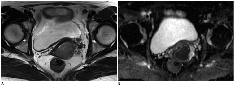
Fig. 1 Method of measurement of ADC values in uterine cervical cancer.Tumor border is identified by visual evaluation on axial T2-weighted image (A). ROI is manually drawn to include tumor as much as possible on ADC map (B) at level corresponding to A. ADC = apparent diffusion coefficient, ROI = region of interest
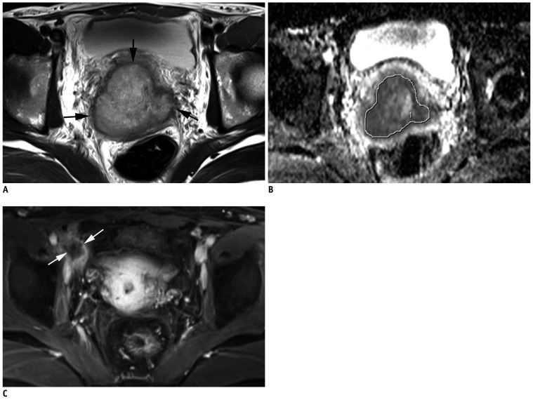
Fig. 2 Thirty-six-year-old woman with tumor recurrence after CRT for advanced uterine cervical cancer.A. Pre-CRT axial T2-weighted image shows uterine cervical cancer (arrows) with left parametrial invasion (FIGO stage IIB). B. Pre-CRT ADC map depicts manually drawn ROI to include tumor as much as possible. ADC75 value of tumor was 1.078 × 10⁻³ mm2/sec. C. Gd-enhanced axial T1-weighted image obtained at 10 months after CRT completion demonstrates about 2 cm metastatic lymphadenopathy (arrows) in right external iliac chain (arrows). ADC = apparent diffusion coefficient, CRT = chemoradiation therapy, FIGO = International Federation of Gynecology and Obstetrics, ROI = region of interest
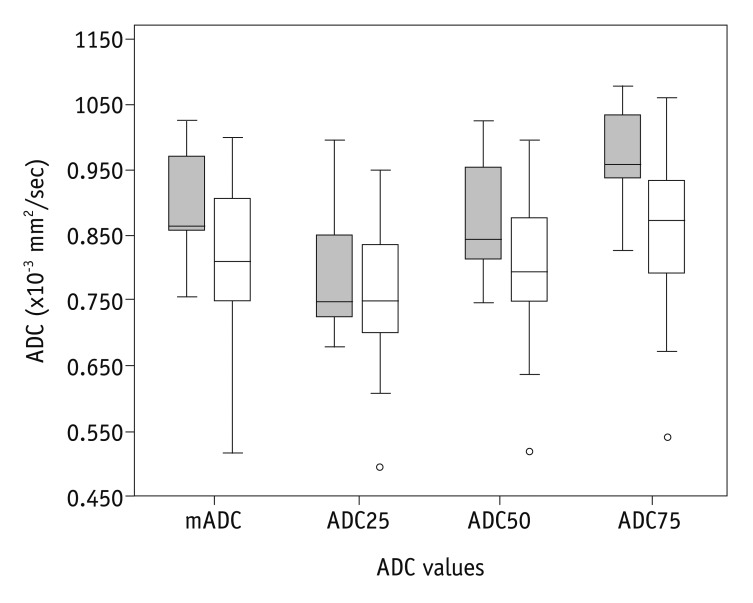
Fig. 3 Boxplots of ADC values of recurrence (shaded box) and no recurrence (open bar) group.Despite substantial overlap between two groups, mADC and ADC75 in recurrence group are significantly higher than those of no recurrence group (p = 0.043 and p = 0.008, respectively). However, ADC25 and ADC50 are not significantly different between two groups (p = 0.087 and p = 0.319, respectively). ADC = apparent diffusion coefficient
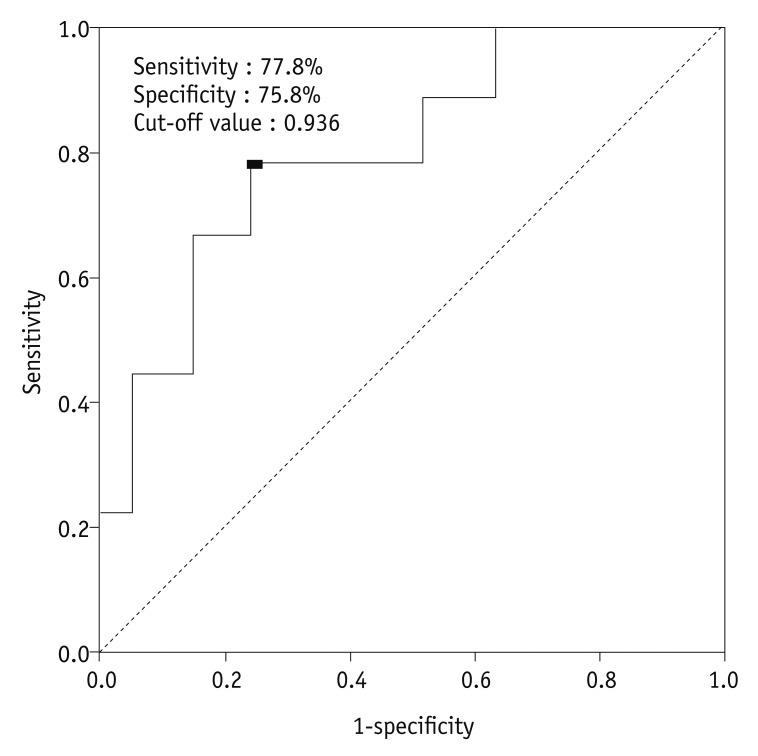
Fig. 4 ROC curve of ADC75 value for predicting tumor recurrence.When cut-off value of ADC75 was 0.936 × 10⁻³ mm2/sec, sensitivity, specificity, and accuracy were 77.8%, 75.8% and 71.1%, respectively. Area under ROC curve (Az) is 0.796 (p = 0.007, 95% CI, 0.636-0.958). ADC = apparent diffusion coefficient, CI = confidence interval, ROC = receiver operating characteristic
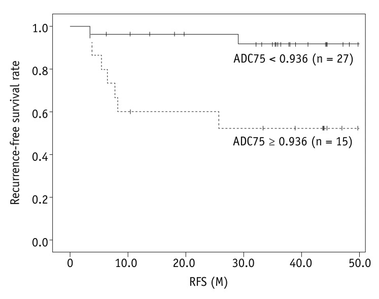
Fig. 5 Recurrence-free survival rate according to ADC75 value.There is statistically significant difference in recurrence-free survival rate between patients with ADC75 ≥ 0.936 × 10⁻³ mm2/sec and ADC75 < 0.936 × 10⁻³ mm2/sec (51.9% vs. 91.7%, p = 0.003, log-rank test). ADC = apparent diffusion coefficient, RFS = recurrence-free survival
Cited by 2 articles
-
Early Prediction of Response to Neoadjuvant Chemotherapy Using Dynamic Contrast-Enhanced MRI and Ultrasound in Breast Cancer
Yunju Kim, Sung Hun Kim, Byung Joo Song, Bong Joo Kang, Kwang-il Yim, Ahwon Lee, Yoonho Nam
Korean J Radiol. 2018;19(4):682-691. doi: 10.3348/kjr.2018.19.4.682.MR Imaging for Staging of Cervical Carcinoma: Update
Seong Kuk Yoon, Dong Won Kim
J Korean Soc Radiol. 2017;77(2):67-84. doi: 10.3348/jksr.2017.77.2.67.
Reference
-
2. Peters WA 3rd, Liu PY, Barrett RJ 2nd, Stock RJ, Monk BJ, Berek JS, et al. Concurrent chemotherapy and pelvic radiation therapy compared with pelvic radiation therapy alone as adjuvant therapy after radical surgery in high-risk early-stage cancer of the cervix. J Clin Oncol. 2000; 18:1606–1613. PMID: 10764420.
Article3. Thomas GM. Improved treatment for cervical cancer--concurrent chemotherapy and radiotherapy. N Engl J Med. 1999; 340:1198–1200. PMID: 10202172.4. Kastritis E, Bamias A, Efstathiou E, Gika D, Bozas G, Zorzou P, et al. The outcome of advanced or recurrent non-squamous carcinoma of the uterine cervix after platinum-based combination chemotherapy. Gynecol Oncol. 2005; 99:376–382. PMID: 16051322.
Article5. Thoeny HC, Ross BD. Predicting and monitoring cancer treatment response with diffusion-weighted MRI. J Magn Reson Imaging. 2010; 32:2–16. PMID: 20575076.
Article6. Heo SH, Jeong YY, Shin SS, Kim JW, Lim HS, Lee JH, et al. Apparent diffusion coefficient value of diffusion-weighted imaging for hepatocellular carcinoma: correlation with the histologic differentiation and the expression of vascular endothelial growth factor. Korean J Radiol. 2010; 11:295–303. PMID: 20461183.
Article7. Lyng H, Haraldseth O, Rofstad EK. Measurement of cell density and necrotic fraction in human melanoma xenografts by diffusion weighted magnetic resonance imaging. Magn Reson Med. 2000; 43:828–836. PMID: 10861877.
Article8. Koh DM, Collins DJ. Diffusion-weighted MRI in the body: applications and challenges in oncology. AJR Am J Roentgenol. 2007; 188:1622–1635. PMID: 17515386.
Article9. Harry VN, Semple SI, Parkin DE, Gilbert FJ. Use of new imaging techniques to predict tumour response to therapy. Lancet Oncol. 2010; 11:92–102. PMID: 20129132.
Article10. Kyriazi S, Collins DJ, Messiou C, Pennert K, Davidson RL, Giles SL, et al. Metastatic ovarian and primary peritoneal cancer: assessing chemotherapy response with diffusion-weighted MR imaging--value of histogram analysis of apparent diffusion coefficients. Radiology. 2011; 261:182–192. PMID: 21828186.
Article11. Charles-Edwards EM, Messiou C, Morgan VA, De Silva SS, McWhinney NA, Katesmark M, et al. Diffusion-weighted imaging in cervical cancer with an endovaginal technique: potential value for improving tumor detection in stage Ia and Ib1 disease. Radiology. 2008; 249:541–550. PMID: 18936313.
Article12. Harry VN, Semple SI, Gilbert FJ, Parkin DE. Diffusion-weighted magnetic resonance imaging in the early detection of response to chemoradiation in cervical cancer. Gynecol Oncol. 2008; 111:213–220. PMID: 18774597.
Article13. Kim HS, Kim CK, Park BK, Huh SJ, Kim B. Evaluation of therapeutic response to concurrent chemoradiotherapy in patients with cervical cancer using diffusion-weighted MR imaging. J Magn Reson Imaging. 2013; 37:187–193. PMID: 23018989.
Article14. Liu Y, Bai R, Sun H, Liu H, Wang D. Diffusion-weighted magnetic resonance imaging of uterine cervical cancer. J Comput Assist Tomogr. 2009; 33:858–862. PMID: 19940650.
Article15. Liu Y, Bai R, Sun H, Liu H, Zhao X, Li Y. Diffusion-weighted imaging in predicting and monitoring the response of uterine cervical cancer to combined chemoradiation. Clin Radiol. 2009; 64:1067–1074. PMID: 19822239.
Article16. McVeigh PZ, Syed AM, Milosevic M, Fyles A, Haider MA. Diffusion-weighted MRI in cervical cancer. Eur Radiol. 2008; 18:1058–1064. PMID: 18193428.
Article17. Naganawa S, Sato C, Kumada H, Ishigaki T, Miura S, Takizawa O. Apparent diffusion coefficient in cervical cancer of the uterus: comparison with the normal uterine cervix. Eur Radiol. 2005; 15:71–78. PMID: 15538578.
Article18. Nakamura K, Joja I, Nagasaka T, Fukushima C, Kusumoto T, Seki N, et al. The mean apparent diffusion coefficient value (ADCmean) on primary cervical cancer is a predictive marker for disease recurrence. Gynecol Oncol. 2012; 127:478–483. PMID: 22892362.
Article19. Somoye G, Harry V, Semple S, Plataniotis G, Scott N, Gilbert FJ, et al. Early diffusion weighted magnetic resonance imaging can predict survival in women with locally advanced cancer of the cervix treated with combined chemo-radiation. Eur Radiol. 2012; 22:2319–2327. PMID: 22653281.
Article20. Kang H, Lee HY, Lee KS, Kim JH. Imaging-based tumor treatment response evaluation: review of conventional, new, and emerging concepts. Korean J Radiol. 2012; 13:371–390. PMID: 22778559.
Article21. Payne GS, Schmidt M, Morgan VA, Giles S, Bridges J, Ind T, et al. Evaluation of magnetic resonance diffusion and spectroscopy measurements as predictive biomarkers in stage 1 cervical cancer. Gynecol Oncol. 2010; 116:246–252. PMID: 19875159.
Article22. Koh DM, Scurr E, Collins D, Kanber B, Norman A, Leach MO, et al. Predicting response of colorectal hepatic metastasis: value of pretreatment apparent diffusion coefficients. AJR Am J Roentgenol. 2007; 188:1001–1008. PMID: 17377036.
Article23. Park SH, Moon WK, Cho N, Song IC, Chang JM, Park IA, et al. Diffusion-weighted MR imaging: pretreatment prediction of response to neoadjuvant chemotherapy in patients with breast cancer. Radiology. 2010; 257:56–63. PMID: 20851939.
Article24. Dzik-Jurasz A, Domenig C, George M, Wolber J, Padhani A, Brown G, et al. Diffusion MRI for prediction of response of rectal cancer to chemoradiation. Lancet. 2002; 360:307–308. PMID: 12147376.
Article25. Zhang Y, Chen JY, Xie CM, Mo YX, Liu XW, Liu Y, et al. Diffusion-weighted magnetic resonance imaging for prediction of response of advanced cervical cancer to chemoradiation. J Comput Assist Tomogr. 2011; 35:102–107. PMID: 21245694.
Article26. Sugahara T, Korogi Y, Kochi M, Ikushima I, Shigematu Y, Hirai T, et al. Usefulness of diffusion-weighted MRI with echo-planar technique in the evaluation of cellularity in gliomas. J Magn Reson Imaging. 1999; 9:53–60. PMID: 10030650.
Article27. Higano S, Yun X, Kumabe T, Watanabe M, Mugikura S, Umetsu A, et al. Malignant astrocytic tumors: clinical importance of apparent diffusion coefficient in prediction of grade and prognosis. Radiology. 2006; 241:839–846. PMID: 17032910.
Article28. Murakami R, Hirai T, Sugahara T, Fukuoka H, Toya R, Nishimura S, et al. Grading astrocytic tumors by using apparent diffusion coefficient parameters: superiority of a one-versus two-parameter pilot method. Radiology. 2009; 251:838–845. PMID: 19318585.29. Pope WB, Kim HJ, Huo J, Alger J, Brown MS, Gjertson D, et al. Recurrent glioblastoma multiforme: ADC histogram analysis predicts response to bevacizumab treatment. Radiology. 2009; 252:182–189. PMID: 19561256.
Article30. Barajas RF Jr, Rubenstein JL, Chang JS, Hwang J, Cha S. Diffusion-weighted MR imaging derived apparent diffusion coefficient is predictive of clinical outcome in primary central nervous system lymphoma. AJNR Am J Neuroradiol. 2010; 31:60–66. PMID: 19729544.
Article31. Harrison LB, Chadha M, Hill RJ, Hu K, Shasha D. Impact of tumor hypoxia and anemia on radiation therapy outcomes. Oncologist. 2002; 7:492–508. PMID: 12490737.
Article32. Vaupel P, Mayer A. Hypoxia in cancer: significance and impact on clinical outcome. Cancer Metastasis Rev. 2007; 26:225–239. PMID: 17440684.
Article33. DeVries AF, Kremser C, Hein PA, Griebel J, Krezcy A, Ofner D, et al. Tumor microcirculation and diffusion predict therapy outcome for primary rectal carcinoma. Int J Radiat Oncol Biol Phys. 2003; 56:958–965. PMID: 12829130.
Article
- Full Text Links
-
- Actions
-
Cited
- CITED
-
- Close
- Share
-
- Similar articles
-
- Diffusion-weighted Magnetic Resonance Imaging for Predicting Response to Chemoradiation Therapy for Head and Neck Squamous Cell Carcinoma: A Systematic Review
- Reversal of a Large Ischemic Lesion with Low Apparent Diffusion Coefficient Value by Rapid Spontaneous Recanalization
- Diffusion-Weighted MR Imaging of Unicystic Odontogenic Tumors for Differentiation of Unicystic Ameloblastomas from Keratocystic Odontogenic Tumors
- Diffusion-weighted MR Imaging after Intracranial Tumor Resection
- Emerging Techniques in Brain Tumor Imaging: What Radiologists Need to Know
