Hepatic Splenosis Preoperatively Diagnosed as Hepatocellular Carcinoma in a Patient with Chronic Hepatitis B: A Case Report
- Affiliations
-
- 1Department of Surgery, Yonsei University College of Medicine, Seoul, Korea. choi5491@yuhs.ac
- 2Department of Internal Medicine, Yonsei University College of Medicine, Seoul, Korea.
- 3Department of Radiology, Yonsei University College of Medicine, Seoul, Korea.
- 4Department of Pathology, Yonsei University College of Medicine, Seoul, Korea.
- KMID: 1713472
- DOI: http://doi.org/10.3346/jkms.2008.23.2.336
Abstract
- We report on a case of hepatic splenosis. A 32-yr-old man underwent a splenectomy due to trauma at the age of 6. He had been diagnosed as being a chronic hepatitis B-virus carrier 16 yr prior to the surgery. The dynamic computer tomography (CT) performed due to elevated serum alpha-fetoprotein (128 ng/mL) demonstrated two hepatic nodules, which were located near the liver capsule. A nodule in Segment IVa had a slight enhancement during both the arterial and portal phases, and another nodule in Segment VI showed a slight enhancement only in the portal phases. Dynamic magnetic resonance imaging (MRI) of the mass in Segment VI showed enhanced development in the arterial phases and slight hyperintensivity to the liver parenchyma in the portal phases. These imaging findings suggested a hypervascular tumor in the liver, which could be either focal nodular hyperplasia, adenoma, or hepatocellular carcinoma (HCC). Even though these lesions were diagnosed as HCC, some of the findings were not compatible with typical HCC. On dynamic CT and MRI, all lesions showed a slight arterial enhancement and did not show early venous washout. All lesions were located near the liver capsule. These findings, along with a history of splenectomy, suggested a diagnosis of hepatic splenosis.
MeSH Terms
-
Adult
Carcinoma, Hepatocellular/complications/*diagnosis/surgery
Focal Nodular Hyperplasia/diagnosis/pathology
Hepatitis B, Chronic/complications/*diagnosis
Humans
Liver/*pathology
Liver Neoplasms/complications/*diagnosis/surgery
Magnetic Resonance Imaging
Male
Splenosis/*diagnosis
Tomography, X-Ray Computed
Treatment Outcome
alpha-Fetoproteins/biosynthesis
Figure
-
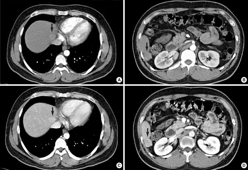
Fig. 1 Arterial phase (A, B) and portal phase (C, D) of abdominal CT scans show a 2-cm mass in Segment IVa and a 3-cm exophytic mass in Segment VI (arrows).
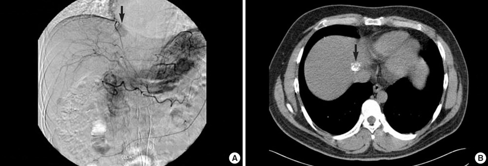
Fig. 2 Hepatic artery angiography shows subtle tumor staining in Segment IVa and no tumor staining in Segment VI (A). A follow-up CT scan taken three weeks later shows lipiodol uptake in the Segment IVa nodule (B).
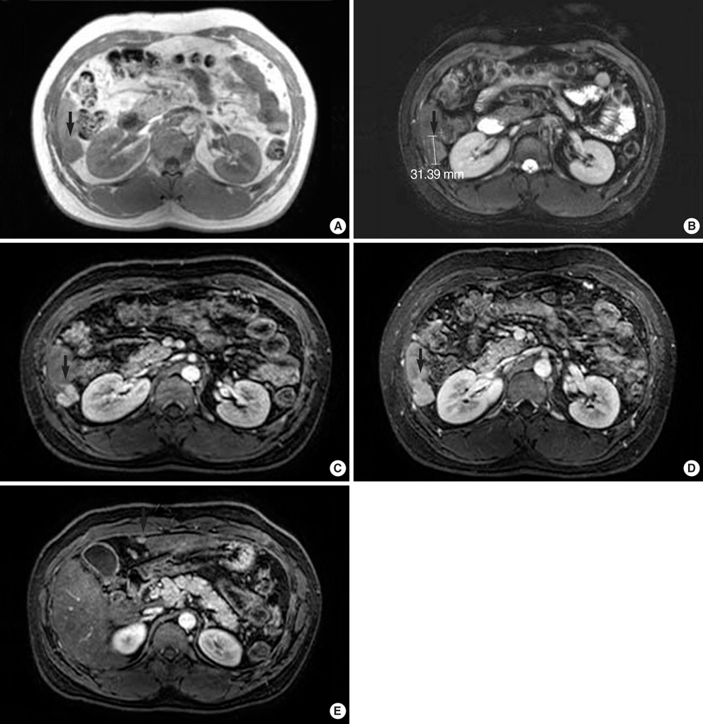
Fig. 3 By precontrast MRI, the lesion in Segment VI shows low signal on T1-weighted (A) and high signal on T2-weighted MR images (B). Dynamic MRI shows an enhancement of the lesion during the arterial phase (C) and a slightly hyperintensive signal in the liver parenchyma during the portal phase (D). Another 1-cm hypervascular mass, which was not detected in previous imaging studies, was found in the inferior tip of Segment IVb (E).
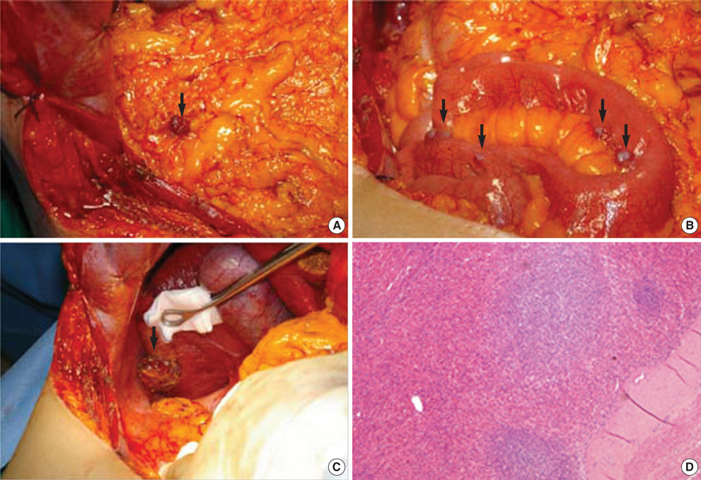
Fig. 4 Photographs of the laparotomy show a 1-cm reddish brown nodule on the greater omentum (A) and small multiple nodules on the surface of the small bowel (B). A 3-cm hepatic nodule on Segment VI was attached to the liver capsule and shows characteristics similar to other abdominal nodules (C). The pathological examination was positive for splenic tissue (H-E stain, ×100) (D).
Cited by 2 articles
-
Thoracic Splenosis: A Case Report and the Importance of Clinical History
Kyungeun Kim, Hye-Jeong Choi, Young Min Kim, Woon Jung Kwon, Won Chan Lee, Jae Hee Suh
J Korean Med Sci. 2010;25(2):299-303. doi: 10.3346/jkms.2010.25.2.299.Intrahepatic Splenosis Mimicking Hepatocellular Carcinoma: A Case Report
Eunjin Hwang, Seonjeong Min, Young Chul Kim, Jung-Ah Choi
J Korean Soc Radiol. 2019;80(2):359-364. doi: 10.3348/jksr.2019.80.2.359.
Reference
-
1. Case records of the Massachusetts General Hospital. Weekly clinicopathological exercises. Case 29-1995. A 65-year-old man with mediastinal Hodgkin's disease and a pelvic mass. N Engl J Med. 1995. 333:784–791.2. Gruen DR, Gollub MJ. Intrahepatic splenosis mimicking hepatic adenoma. AJR Am J Roentgenol. 1997. 168:725–726.
Article3. Yoshimitsu K, Aibe H, Nobe T, Ezaki T, Tomoda H, Hayashi I, Koga M. Intrahepatic splenosis mimicking a liver tumor. Abdom Imaging. 1993. 18:156–158.
Article4. Lee JB, Ryu KW, Song TJ, Suh SO, Kim YC, Koo BH, Choi SY. Hepatic splenosis diagnosed as hepatocellular carcinoma: report of a case. Surg Today. 2002. 32:180–182.
Article5. De Vuysere S, Van Steenbergen W, Aerts R, Van Hauwaert H, Van Beckevoort D, Van Hoe L. Intrahepatic splenosis: imaging features. Abdom Imaging. 2000. 25:187–189.
Article6. Di Costanzo GG, Picciotto FP, Marsilia GM, Ascione A. Hepatic splenosis misinterpreted as hepatocellular carcinoma in cirrhotic patients referred for liver transplantation: report of two cases. Liver Transpl. 2004. 10:706–709.
Article7. Brancatelli G, Vilgrain V, Zappa M, Lagalla R. Case 80: splenosis. Radiology. 2005. 234:728–732.
Article8. Kondo M, Okazaki H, Takai K, Nishikawa J, Ohta H, Uekusa T, Yoshida H, Tanaka K. Intrahepatic splenosis in a patient with chronic hepatitis C. J Gastroenterol. 2004. 39:1013–1015.
Article9. D'Angelica M, Fong Y, Blumgart LH. Isolated hepatic splenosis: first reported case. HPB Surg. 1998. 11:39–42.10. Zhao M, Xu HW. Splenosis simulating an intrahepatic mass. Chin J Traumatol. 2004. 7:62–64.11. Massey MD, Stevens JS. Residual spleen found on denatured red blood cell scan following negative colloid scans. J Nucl Med. 1991. 32:2286–2287.
- Full Text Links
-
- Actions
-
Cited
- CITED
-
- Close
- Share
-
- Similar articles
-
- Intrahepatic Splenosis Mimicking Hepatocellular Carcinoma: A Case Report
- A Case of Composite Hepatocellular Carcinoma and Neuroendocrine Carcinoma in a Patient with Liver Cirrhosis Caused by Chronic Hepatitis B
- Primary Hepatic Tuberculosis Mimicking Hepatocelluar Carcinoma in Patient with Chronic Viral Hepatitis B and C
- Splenosis Mimicking Hepatocellular Carcinoma
- Quantitative hepatitis B surface antigen predicts the antiviral response and hepatocellular carcinoma development in patients with chronic hepatitis B
