J Korean Med Sci.
2007 Feb;22(1):153-155. 10.3346/jkms.2007.22.1.153.
Gastric Accumulation of Bone Seeking Agent in a Patient with Advanced Gastric Cancer
- Affiliations
-
- 1Department of Nuclear Medicine, Wonkwang University School of Medicine, Iksan, Korea. akaxan@nate.com
- 2Department of Nuclear Medicine and Research Institute of Clinical Medicine, Chonbuk National University Medical School, Jeonju, Korea.
- KMID: 1713257
- DOI: http://doi.org/10.3346/jkms.2007.22.1.153
Abstract
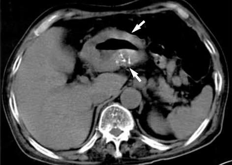
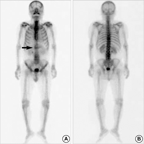
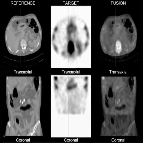
- Soft tissue uptake of Tc-99m labeled bone seeking agents, such as Tc-99m 3,3-diphosphono-1,2-propanedicarboxylic acid (DPD), is commonly seen in clinical practice, even though bone scintigraphy is mainly used to detect bone disease. However, gastric uptake of bone agents in patients with gastric cancer is very rare. And it has been reported that calcified gastric adenocarcinoma appears in only about 5% of all gastric cancer. We report a rare case of bone scintigraphy, single photon emission computed tomography and computed tomography fusion images that demonstrated diffuse gastric uptake of Tc-99m DPD in a patient with advanced gastric cancer.
MeSH Terms
Figure
Reference
-
1. Rotondo A, Grassi R, Smaltino F, Greco A, Leung AW, Allison DJ. Calcified gastric cancer: report of a case and review of the literature. Br J Radiol. 1986. 59:405–407.2. Balestreri L, Canzonieri V, Morassut S. Calcified gastric cancer-CT findings before and after chemotherapy. Case report and discussion of the pathogenesis of this type of calcification. Clin Imaging. 1997. 21:122–125.
Article3. Peller PJ, Ho VB, Kransdorf MJ. Extraosseous Tc-99m MDP uptake: a pathophysiologic approach. Radiographics. 1993. 13:715–734.
Article4. Gruber GB. Knochebildung in einem magen karzinom. Z Beitr Path Anat. 1913. 55:368–370.5. Kunieda K, Okuhira M, Nakano T, Nakatani S, Tateiwa J, Hiramatsu A, Mizuno T, Shiozaki Y, Sameshima Y. Diffuse calcification in gastric cancer. J Int Med Res. 1990. 18:506–514.
Article6. De Carvalho JC, Francischetti EA, De Barros Filho GA, Cerda JJ. Calcified mucinous adenocarcinoma of the stomach. Am J Gastroenterol. 1978. 69:481–484.7. Adachi Y, Mori M, Kido A, Shimono R, Maehara Y, Sugimachi K. A clinicopathologic study of mucinous gastric carcinoma. Cancer. 1992. 69:866–871.
Article8. Ashley DJB. Evan's Histological Appearances of Tumors. 1990. Edinburgh: Churchill Livingstone;307–308.9. Delcourt E, Baudoux M, Neve P. Tc-99m-MDP bone scanning detection of gastric calcification. Clin Nucl Med. 1980. 5:546–547.
Article10. Watanabe N, Haida M, Mikuni I, Shimizu M, Nakajima A, Seto H. Bone imaging in advanced gastric cancer. Clin Nucl Med. 1998. 23:384.
Article
- Full Text Links
- Actions
-
Cited
- CITED
-
- Close
- Share
- Similar articles
-
- Neoadjuvant Chemotherapy in Asian Patients With Locally Advanced Gastric Cancer
- Bone Scanning in the Evaluation of Lung Cancer
- Role of Gastric Stem Cells in Gastric Carcinogenesis by Chronic Helicobacter pylori Infection
- Corrigendum: Korean Gastric Cancer Association Nationwide Survey on Gastric Cancer in 2014
- Surgical Outcomes of Patients Undergoing Gastrectomy for Gastric Cancer: Does the Age Matter?


