Cutaneous Metastasis of Choriocarcinoma: A Case Report
- Affiliations
-
- 1Department of Dermatology, Yonsei University College of Medicine, Seoul, Korea.
- 2Department of Pathology, Yonsei University College of Medicine, Seoul, Korea. dbang@yumc.yonsei.ac.kr
- KMID: 1712756
- DOI: http://doi.org/10.3346/jkms.2005.20.4.683
Abstract
- Choriocarcinoma is one of the malignant tumors of trophoblastic cells characterized by the secretion of human chorionic gonadotrophin (hCG) (1-3). Cutaneous metastasis is a rare presentation of choriocarcinoma but a poor prognostic sign because it is associated only with widespread disease (3-5). A 52-yr-old female complaining of dyspnea for 2 months, presented with fingertip sized erythematous nodules on the left side of the neck and the right side of the upper back of 1 month duraton. She has suffered from Behcet's disease since 1999. Microscopic examination of a nodule of upper back demonstrated biphasic pattern of cytotrophoblasts and hCG-positive syncytiotrophoblasts, and the typical histologic features of choriocarcinoma. She was referred to the gynecological oncology department. After 17 cycles of combination chemotherapy, the serum hCG level has fallen from 700,000 to under 2.0 mIU/mL and the skin lesions have almost disappeared. However, after 3 months, total abdominal hysterectomy and bilateral salpingo-oophorectomy were performed due to elevated serum hCG level (4,447.7 mIU/mL), and she is scheduled to receive post-operative adjuvant chemotherapy.
Keyword
MeSH Terms
Figure
-
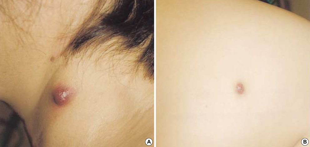
Fig. 1 Fingernail sized erythematous nodules. (A) left side of neck, (B) right side of upper back.
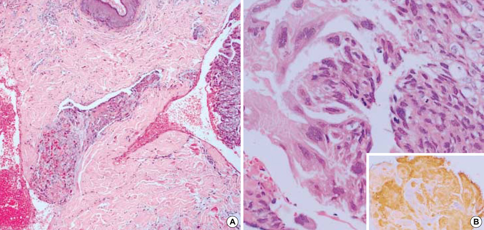
Fig. 2 (A) Several hemorrhagic foci and multiple cellular infiltration throughout the dermis (hematoxylin and eosin, ×40). (B) Infiltrated tumor cells are composed of two populations of cells. One element exhibits clear cytoplasm and large, vesicular nuclei with prominent nucleoli, which is identified as cytotrophoblasts. The other cell population shows eosinophilic cytoplasm with cytoplasmic vacuoles and many dark nuclei, which is identified as syncytiotrophoblasts (hematoxylin and eosin, ×200). (Inset) hCG was positively stained in the cytoplasm of syncytiotrophoblasts but not the cytotrophoblasts (hCG stain, ×400).

Fig. 3 Abdomen pelvis MR images shows a protruding mass from low uterine cavity to vaginal canal (short arrow) and diffuse enlargement of uterus body showing heterogenous signal intensity, suggesting myometrial invasion (long arrow).
Cited by 1 articles
-
Cutaneous Metastasis of Choriocarcinoma
Jaewoong Choi, Noo Ri Lee, Jin Wook Lee, Sung Ku Ahn
Ann Dermatol. 2015;27(6):793-794. doi: 10.5021/ad.2015.27.6.793.
Reference
-
1. Copeland LJ. Copeland LJ, Jarrell JF, editors. Gestational trophoblastic neoplasia. Textbook of gynecology. 2000. 2nd ed. Philadelphia: W.B. Saunders;1418–1427.2. Clement PB, Young RH. Clement PB, Young RH, editors. Ovarian germ cell tumors. Atlas of gynecologic surgical pathology. 2000. Philadelphia: W.B. Saunders;346.3. Shimizu S, Nagata Y, Han-yaku H. Metastatic testicular choriocarcinoma of the skin: Report and review of the literature. Am J Dermatopathol. 1996. 18:633–636.4. Chhieng DC, Jennings TA, Slominski A, Mihm MC Jr. Choriocarcinoma presenting as a cutaneous metastasis. J Cutan Pathol. 1995. 22:374–377.
Article5. Chama CM, Nggada HA, Nuhu A. Cutaneous metastasis of gestational choriocarcinoma. Int J Gynaecol Obstet. 2002. 77:249–250.
Article6. Johnson WC. Elder D, Elenitsas R, Jaworsky C, editors. Metastatic carcinoma of the skin. Lever's histopathology of the skin. 1997. Philadelphia: Lippincott-Raven;1011–1018.7. Odom RB, James WD, Berger TG, editors. Metastatic carcinoma. Andrew's diseases of the skin. 2000. 9th ed. Philadelphia: W.B. Saunders;794–797.8. Brownstein MH, Helwig EB. Patterns of cutaneous metastasis. Arch Dermatol. 1972. 105:862–868.
Article9. Spencer PS, Helm TN. Skin metastases in cancer patients. Cutis. 1987. 39:119–121.10. Lookingbill DP, Spangler N, Sexton FM. Skin involvement as the presenting sign of internal carcinoma. J Am Acad Dermatol. 1990. 22:19–26.
Article11. Lookingbill DP, Spangler N, Helm KF. Cutaneous metastases in patients with metastatic carcinoma: A retrospective study of 4020 patients. J Am Acad Dermatol. 1993. 29:228–236.
Article
