The Usefulness of CDX-2 for Differentiating Primary and Metastatic Ovarian Carcinoma: An Immunohistochemical Study Using a Tissue Microarray
- Affiliations
-
- 1Department of Pathology, College of Medicine, Yeungnam University, Daegu, Korea. mjkap@med.yu.ac.kr
- KMID: 1712747
- DOI: http://doi.org/10.3346/jkms.2005.20.4.643
Abstract
- Distinguishing primary ovarian carcinoma from metastatic carcinoma to the ovary is often difficult by histologic examination alone. Recently an immunohistochemical marker CDX-2 was found to be of considerable diagnostic value in establishing the gastrointestinal origin of metastatic tumors. The aim of this study was to determine whether CDX-2 can distinguish between these malignancies. Paraffin-embedded tissue sections from 57 primary ovarian tumors and 40 metastatic tumors to the ovary were immunostained for CDX-2, and results were compared to the ancillary immunohistochemical results for CK7/CK20, CEA, CA125, and her-2/neu. CDX-2 immunoreactivity was observed in most of metastatic carcinomas with colorectal (91%) and appendiceal (100%) origin, however CDX-2 was negative in all primary ovarian carcinomas, except for the mucinous subtype. Almost all primary ovarian carcinomas including the mucinous subtype showed diffuse and strong immunoexpression for CK7. CEA and CA125 were mainly found in metastatic and primary ovarian carcinoma, respectively. Her-2/neu overexpression was only noted in a small proportion of primary and metastatic ovarian carcinomas. These results suggest that CDX-2 is very useful immunohistochemical marker for distinguishing metastatic colorectal carcinoma to the ovary from primary ovarian carcinoma, including the mucinous subtype. Furthermore, combination with CDX-2 and CK7 strengthen the differential diagnosis between these tumors.
Keyword
MeSH Terms
-
CA-125 Antigen/analysis
Carcinoembryonic Antigen/analysis
Colorectal Neoplasms/metabolism/pathology
Diagnosis, Differential
Female
Homeodomain Proteins/*analysis
Humans
Immunohistochemistry
Keratin/analysis
Neoplasm Metastasis
Ovarian Neoplasms/metabolism/*pathology
Receptor, erbB-2/analysis
Research Support, Non-U.S. Gov't
Tissue Array Analysis/*methods
Trans-Activators/*analysis
Figure
-

Fig. 1 Representative sections of diffuse CDX-2 immunoreactivity in primary ovarian mucinous adenocarcinoma (A) and in metastatic carcinoma from colorectum (B), stomach (C) and appendix (D) (×40).
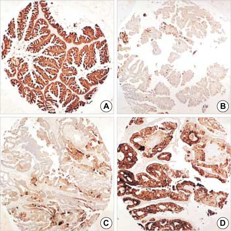
Fig. 2 Almost all primary ovarian mucinous carcinomas are diffusely positive for CK7 (A), and variably positive for CK20, often showing patchy rather than diffuse staining (B). Conversely, metastatic colorectal carcinoma had a focal CK7 (C) and diffuse CK20 (D) immunoprofile (×40).
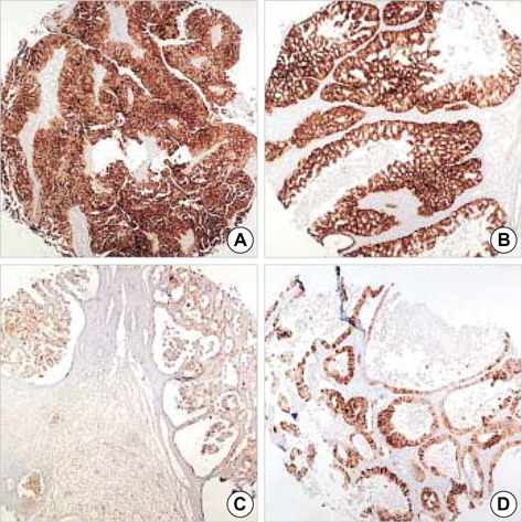
Fig. 3 Diffuse strong cytoplasmic staining for CA125 is observed in ovarian serous carcinoma (A). Her-2/neu expression is evident in a small group of the ovarian mucinous (B) and clear cell carcinomas (C) and unexpectidly in metastatic carcinomas from the stomach (D) (×40).
Reference
-
1. Scully RE, Young RH, Philip BC. Atlas of tumor pathology: Tumors of the ovary, maldeveloped gonads, fallopian tube, and broad ligament. 1998. Washington D.C: AFIP;3rd series.2. Daya D, Nazerali L, Frank GL. Metastatic ovarian carcinoma of large intestinal origin simulating primary ovarian carcinoma. A clinicopathologic study of 25 cases. Am J Clin Pathol. 1992. 97:751–758.
Article3. Ueda G, Sawada M, Ogawa H, Tanizawa O, Tsujimoto M. Immunohistochemical study of cytokeratin-7 for the differential diagnosis of adenocarcinomas in the ovary. Gynecol Oncol. 1993. 51:219–223.4. Loy TS, Calaluce RD, Keeney GL. Cytokeratin immunostaining in differentiating primary ovarian carcinoma from metastatic colonic adenocarcinoma. Mod Pathol. 1996. 9:1040–1044.5. Berezowski K, Stastny JF, Kornstein MJ. Cytokeratin 7 and 20 and carcinoembryonic antigen in ovarian and colonic carcinoma. Mod Pathol. 1996. 9:426–429.6. Fowler LJ, Maygarden SJ, Novotny DB. Human alveolar macrophage-56 and carcinoembryonic antigen monoclonal antibdies in the differential diagnosis between primary ovarian and metastatic gastrointestinal carcinomas. Hum Pathol. 1994. 25:666–670.7. Albarracin CT, Jafri J, Montag AG, Hart J, Kuan SF. Differential expression of MUC2 and MUC5AC mucin genes in primary ovarian and metastatic colonic carcinoma. Hum Pathol. 2000. 31:672–677.8. Suh E, Traber PG. An intestine-specific homeobox gene regulates proliferation and differentiation. Mol Cell Biol. 1996. 16:619–625.
Article9. Seno H, Oshima M, Taniguchi MA, Usami K, Ishikawa TO, Chiba T, Taketo MM. CDX2 expression in the stomach with intestinal metaplasia and intestinal-type cancer: prognostic implications. Int J Oncol. 2002. 21:769–774.
Article10. Werling RW, Yaziji H, Bacchi CE, Gown AM. CDX2, a highly sensitive and specific marker of adenocarcinomas of intestinal origin: an immunohistochemical survey of 476 primary and metastatic carcinomas. Am J Surg Pathol. 2003. 27:303–310.11. Lee KR, Young RH. The distinction between primary and metastatic mucinous carcinomas of the ovary: gross and histologic findings in 50 cases. Am J Surg Pathol. 2003. 27:281–292.12. Kononen J, Bubendorf L, Kallioniemi A, Barlund M, Schraml P, Leighton S, Torhorst J, Mihatsch MJ, Sauter G, Kallioniemi OP. Tissue microarrays for high-throughput molecular profiling of tumor specimens. Nat Med. 1998. 4:844–847.
Article13. Bai YQ, Yamamoto H, Akiyama Y, Tanaka H, Takizawa T, Kake M, Kenji Yagi O, Saitoh K, Takeshita K, Iwai T, Yuasa Y. Ectopic expression of homeodomain protein CDX2 in intestinal metaplasia and carcinomas of the stomach. Cancer Lett. 2002. 176:47–55.
Article14. Li MK, Folpe AL. CDX-2, a new marker for adenocarcinom of gastrointestinal origin. Adv Anat Pathol. 2004. 11:101–105.15. Eda A, Osawa H, Yanaka I, Satoh K, Mutoh H, Kihira K, Sugano K. Expression of homeobox gene CDX2 precedes that of CDX1 during the progression of intestinal metaplasia. J Gastroenterol. 2002. 37:94–100.
Article16. Vassil K, Luig T, Luig T. The homeobox intestinal differentiation factor CDX2 is selectively expressed in gastrointestinal adenocarcinomas. Mod Pathol. 2004. 17:1392–1399.
Article17. Groisman GM, Meir A, Sabo E. The value of CDX2 immunostaining in differentiating primary ovarian carcinomas from colonic carcinomas metastatic to the ovaries. Int J Gynecol Pathol. 2004. 23:52–57.
Article18. Logani S, Oliva E, Arnell PM, Amin MB, Young RH. Use of novel immunohistochemical markers expressed in colonic adenocarcinoma to distinguish primary ovarian tumors from metastatic colorectal carcinoma. Mod Pathol. 2005. 18:19–25.
Article19. Fraggetta F, Peloci G, Cafici A, Nuciforo P, Viale G. CDX2 immunoreactivity in primary and metastatic ovarian mucinous tumors. Virchows Arch. 2003. 443:782–786.20. Kaimaktchieve V, Terracciano L, Tornillo L, Spichtin H, Stoios D, Bundi M, Korcheva V, Mirlacher M, Loda M, Sauter G, Corless CL. The homeobox intestinal differentiation factor CDX 2 is selectively expressed in gastrointestinal adenocarcinomas. Mod Pathol. 2004. 17:1392–1399.21. Barbareschi M, Murer B, Colby TV, Chilosi M, Macri E, Loda M, Doglioni C. CDX-2 homeobox gene expression is a reliable marker of colorectal adenocarcinoma metastatses to the lungs. Am J Surg Pathol. 2003. 27:141–149.22. Raspollini MR, Baroni G, Taddei A, Taddei GL. Primary cervical adenocarcinoma with intestinal differentiation and colonic carcinoma metastatic to cervix. An investigation using CDX-2 and a limited immunohistochemical panel. Arch Pathol Lab Med. 2003. 127:1586–1590.
Article23. Franchi A, Massi D, Palomba A, Biancalani M, Santucci M. CDX-2, cytokeratin 7 and cytokeratin 20 immunohistochemical expression in the differential diagnosis of primary adenocarcinomas of the sinonasal tract. Virchows Arch. 2004. 445:63–67.
Article24. McCluggage WG. Recent advances in immunohistochemistry in the diagnosis of ovarian neoplasm. J Clin Pathol. 2000. 53:327–334.25. Lagendijk JH, Mullink H, Van Diest PJ, Meijer GA, Meijer CJ. Tracing the origin of adenocarcinomas with unknown primary using immunohistochemistry: differential diagnosis between colonic and ovarian carcinomas as primary sites. Hum Pathol. 1998. 29:491–497.
Article26. Wauters CC, Smedts F, Gerrits LG, Bosman FT, Ramaekers FC. Keratins 7 and 20 as a dignostic markers of carcinomas metastatic to the ovary. Hum Pathol. 1995. 26:852–855.27. Lash RH, Hart WR. Intestinal adenocarcinomas metastatic to the ovaries. A clinicopathologic evaluation of 22 cases. Am J Surg Pathol. 1987. 11:114–121.28. Lagendijk JH, Mullink H, van Diest PJ, Meijer GA, Meijer CJ. Immunohistochemical differentiation between primary adenocarcinomas of the ovary and ovarian metastases of colonic and breast origin. Comparison between a statistical and an intuitive approach. J Clin Pathol. 1999. 52:283–290.
Article29. Cirisano FD, Karlan BY. The role of the HER-2/neu oncogene in gynecologic cancers. J Soc Gynecol Invest. 1996. 3:99–105.
Article30. Iwamoto H, Fukasawa H, Honda T, Hirata S, Hoshi K. HER-2/neu expression in ovarian clear cell carcinomas. Int J Gynecol Cancer. 2003. 13:28–31.
Article
- Full Text Links
-
- Actions
-
Cited
- CITED
-
- Close
- Share
-
- Similar articles
-
- Immunohistochemical Studies for Differential Diagnosis between Primary and Metastatic OvarianEpithelial Tumors
- Perimenopausal Ovarian Carcinoma Patient with Subclavian Node Metastasis Proven by Immunohistochemistry
- The Usefulness of Cytokeratin 7 and Colon Ovarian Tumor Antigen in the Differential Diagnosis of Primary and Metastatic Ovarian Tumors
- Ovarian and Pituitary Metastasis from Adenocarcinoma of the Lung: A Case Report
- CDX-1/CDX-2 Expression Is a Favorable Prognostic Factor in Epstein-Barr Virus-Negative, Mismatch Repair-Proficient Advanced Gastric Cancers
