Multi-Detector Row Computed Tomographic Evaluation of a Rare Type of Complete Vascular Ring: Double Aortic Arch with Atretic Left Arch Distal to the Origin of Left Subclavian Artery
- Affiliations
-
- 1Department of Radiology, Taichung Veterans General Hospital, Taichung 407, Taiwan. sillyduck.radiology@gmail.com
- 2Section of Pediatric Cardiology, Department of Pediatrics, Taichung Veterans General Hospital, Taichung 407, Taiwan.
- 3Section of Cardiovascular Surgery, Cardiovascular Center, Taichung Veterans General Hospital, Taichung 407, Taiwan.
- 4Department of Medical Imaging, Show Chwan Memorial Hospital and Chang Bing Show Chwan Memorial Hospital, Changhua 500, Taiwan.
- 5Department of Radiological Technology and Graduate Institute of Radiological Science, Central Taiwan University of Science and Technology, Taichung 406, Taiwan.
- KMID: 1711445
- DOI: http://doi.org/10.3348/kjr.2013.14.5.845
Abstract
- Double aortic arch with an atretic left arch distal to the origin of left subclavian artery was diagnosed with multi-detector row computed tomography (MDCT) in two children with dysphagia. This rare type of complete vascular ring is clinically important because it may be confused with right aortic arch in mirror imaging. Anatomic details of this rare type of complete vascular ring demonstrated on MDCT facilitated appropriate surgical treatment.
MeSH Terms
-
Adolescent
Aorta, Thoracic/*abnormalities/radiography/surgery
Child, Preschool
Deglutition Disorders/etiology/radiography/surgery
Female
Humans
Multidetector Computed Tomography/*methods
Subclavian Artery/*abnormalities/radiography/surgery
Vascular Malformations/complications/*radiography/surgery
Vascular Surgical Procedures
Figure
-
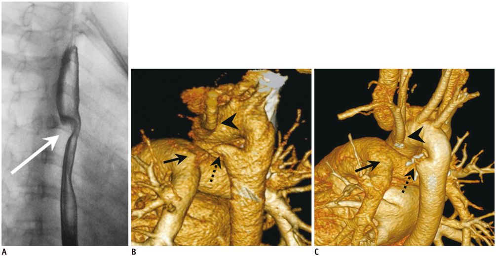
Fig. 1 Key medicl images of case 1. A. Double contrast esophagogram of Case 1. Left posterior oblique view of esophagus revealed posterior indentation over middle portion (arrow). Pre- and post-operational multi-detector row computed tomography images of Case 1. B. Posterior volume rendering image shows proximity of posterioinferiorly distorted LSCA (arrowhead), descending aortic diverticulum (dashed arrow), and pulmonary artery pouch (arrow) before surgery. C. Posterior volume rendering image one year after division and oversewing. Upward migration of LSCA (arrowhead) and separation of descending aorta diverticulum (dashed arrow) and pulmonary artery pouch (arrow) are shown. LSCA = left subclavian artery
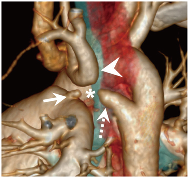
Fig. 2 Multi-detector row computed tomography image of Case 2. Posterior volume rendering image. Proximity of posteroinferiorly distorted left subclavian artery (arrowhead), descending aortic diverticulum (dashed arrow) and pulmonary artery pouch (arrow) suggests fibrous connection (*) among these structures, and demonstrates double aortic arch with atretic left aortic arch distal to origin of left subclavian artery.
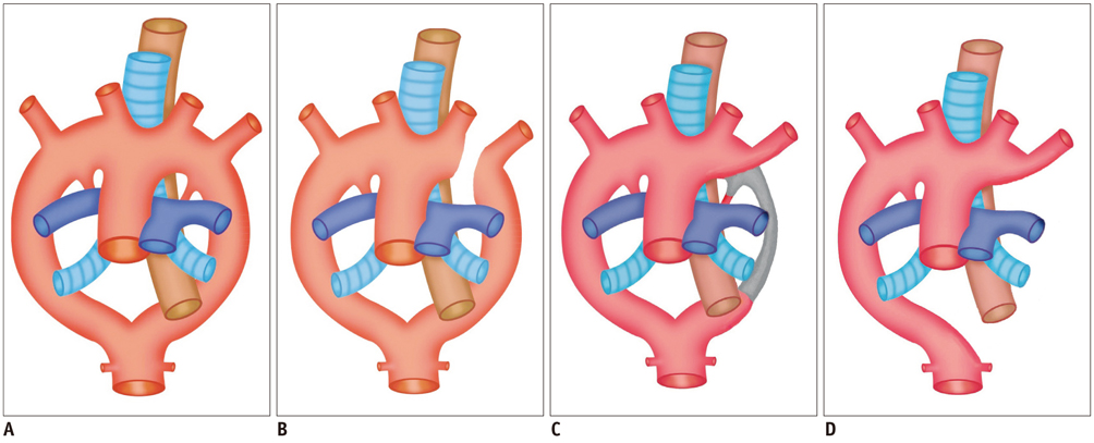
Fig. 3 Schematic figure of vascular ring. A. Double aortic arch is one of most common causes of vascular ring. B. Right aortic arch with aberrant left subclavian artery from diverticulum of Kommerell also results in vascular ring. C. Double aortic arch with atretic left aortic arch distal to origin of left subclavian artery is rare type of vascular ring. D. Right aortic arch with mirror image branching does not comprise vascular ring, but mimics vascular ring type of double aortic arch with atretic left aortic arch distal to origin of left subclavian artery (C).
Reference
-
1. Tsai IC, Lee T, Chen MC, Tsai WL, Lin PC, Liao WC. Homogeneous enhancement in pediatric thoracic CT aortography using a novel and reproducible method: contrast-covering time. AJR Am J Roentgenol. 2007; 188:1131–1137.2. Schlesinger AE, Krishnamurthy R, Sena LM, Guillerman RP, Chung T, DiBardino DJ, et al. Incomplete double aortic arch with atresia of the distal left arch: distinctive imaging appearance. AJR Am J Roentgenol. 2005; 184:1634–1639.3. Holmes KW, Bluemke DA, Vricella LA, Ravekes WJ, Kling KM, Spevak PJ. Magnetic resonance imaging of a distorted left subclavian artery course: an important clue to an unusual type of double aortic arch. Pediatr Cardiol. 2006; 27:316–320.4. Hong YT, Fu YC, Chen CH, Jan SL, Wang TM, Chang Y, et al. Vascular ring due to double aortic arch with atretic left arch and left ligamentum arteriosum: report of one case. Acta Paediatr Taiwan. 2003; 44:168–170.5. Kellenberger CJ. Aortic arch malformations. Pediatr Radiol. 2010; 40:876–884.6. Edwards JE. Anomalies of the derivatives of the aortic arch system. Med Clin North Am. 1948; 32:925–949.7. Türkvatan A, Büyükbayraktar FG, Olçer T, Cumhur T. Congenital anomalies of the aortic arch: evaluation with the use of multidetector computed tomography. Korean J Radiol. 2009; 10:176–184.
- Full Text Links
-
- Actions
-
Cited
- CITED
-
- Close
- Share
-
- Similar articles
-
- Anomalous Origins of the Bilateral Vertebral Arteries Arising from the Aortic Arch: A Case Report
- Two Cases of Aberrant Vertebral Artery Originating from Aortic Arch Distal to Left Subclavian Artery
- Surgical Correction of Complete Vascular Ring Associated with Kommerell's Diverticulum
- Right Aortic Arch with a Retroesophageal Left Subclavian Artery and an Anomalous Origin of the Pulmonary Artery from the Aorta
- Right-sided aortic arch with the retroesophageal left subclavian artery as the fourth branch
