Primary Malt Lymphoma of the Common Bile Duct
- Affiliations
-
- 1Department of Radiology and Research Institute of Radiology, University of Ulsan College of Medicine, Asan Medical Center, Seoul 138-736, Korea. jhbyun@amc.seoul.kr
- KMID: 1711430
- DOI: http://doi.org/10.3348/kjr.2013.14.5.764
Abstract
- Primary mucosa-associated lymphoid tissue (MALT) lymphoma arising in the common bile duct (CBD) is extremely rare. In our case of MALT lymphoma, CT and MRI showed long, segmental, irregular wall thickening of the CBD and minimal dilatation of the upstream bile duct. A preoperative diagnosis of cholangiocarcinoma was made, but histologic evaluation confirmed MALT lymphoma of the CBD. We herein present a rare case of MALT lymphoma of the CBD with CT and MRI findings.
MeSH Terms
Figure
-
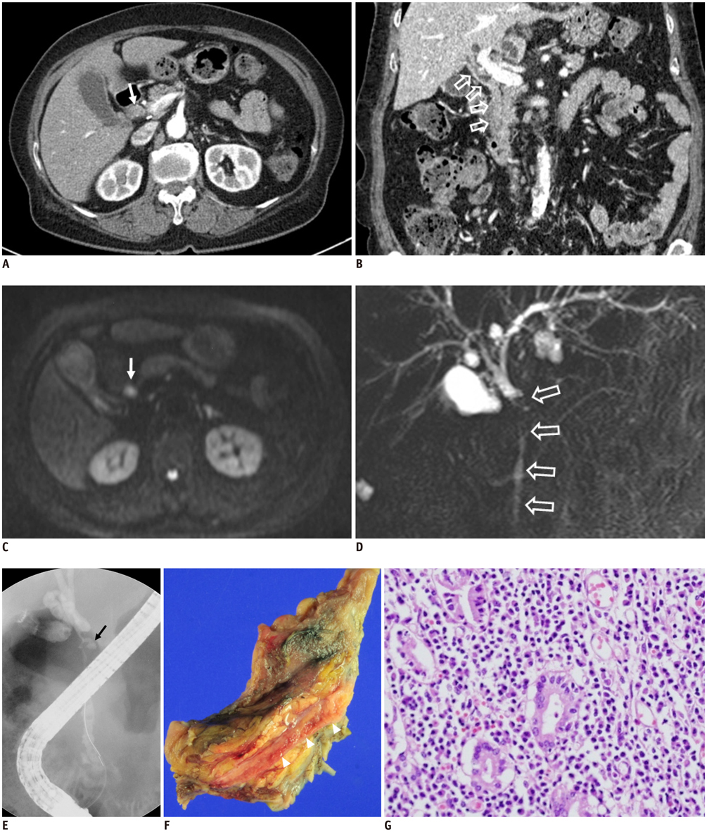
Fig. 1 Seventy nine-year-old male patient with abdominal pain and progressive jaundice. A. CT scan of abdomen during arterial phase reveals concentric wall thickening of common bile duct (CBD; arrow) with similar degree of enhancement compared to hepatic parenchyma. B. Coronal reconstructed CT image during portal phase shows long segmental, irregular wall thickening (open arrows) of CBD. C. Diffusion-weighted MR imaging (b-value of 800) demonstrates heterogeneous high signal intensity (arrow) of thickened wall of CBD. D. MR cholangiopancreatography using single-shot rapid acquisition with relaxation enhancement sequence (TR/TE, infinite/1000; echo train length, 256; FOV, 250 × 250; slice thickness, 40 mm) shows irregular luminal narrowing of entire CBD (open arrows) and mild dilatation of common hepatic duct and intrahepatic duct. Incidental pancreatic divisum is also noted. TR = time to repetition, TE = echo time, FOV = field of view. E. Endoscopic retrograde cholangiopancreatography reveals diffuse narrowing and irregularity of CBD lumen with mild dilatation of upstream intrahepatic bile ducts. Small, saccular out-pouching lesion (arrow) is also noted in proximal CBD. F. Photography for gross specimen reveals diffuse thickening of CBD wall (arrow heads). G. Histologic examination under light microscopy reveals dense and diffuse infiltration of atypical lymphoid cells within CBD walls as well as formation of lymphoid follicles (H and E staining, × 200). CBD = common bile duct
Cited by 1 articles
-
Primary Biliary Mucosa-associated Lymphoid Tissue Lymphoma Mimicking Hilar Cholangiocarcinoma
Seungha Hwang, Tae Jun Song, Seol So, Min Kyung Jeon, Eun Hye Oh, Byoung Soo Kwon, Sujong An, Myung-Hwan Kim
Korean J Gastroenterol. 2016;68(2):114-118. doi: 10.4166/kjg.2016.68.2.114.
Reference
-
1. Matasar MJ, Zelenetz AD. Overview of lymphoma diagnosis and management. Radiol Clin North Am. 2008; 46:175–198. vii2. Isaacson P, Wright DH. Malignant lymphoma of mucosa-associated lymphoid tissue. A distinctive type of B-cell lymphoma. Cancer. 1983; 52:1410–1416.3. Boccardo J, Khandelwal A, Ye D, Duke BE. Common bile duct MALT lymphoma: case report and review of the literature. Am Surg. 2006; 72:85–88.4. Hayashi D, Devenney-Cakir B, Lee JC, Kim SH, Cheng J, Goldfeder S, et al. Mucosa-associated lymphoid tissue lymphoma: multimodality imaging and histopathologic correlation. AJR Am J Roentgenol. 2010; 195:W105–W117.5. Hwang DW, Lim CS, Jang JY, Lee SE, Yoon SO, Jeon YK, et al. Primary hematolymphoid malignancies involving the extrahepatic bile duct or gallbladder. Leuk Lymphoma. 2010; 51:1278–1287.6. Kang CS, Lee YS, Kim SM, Kim BK. Primary low-grade B cell lymphoma of mucosa-associated lymphoid tissue type of the common bile duct. J Gastroenterol Hepatol. 2001; 16:949–951.7. Yoon MA, Lee JM, Kim SH, Lee JY, Han JK, Choi BI, et al. Primary biliary lymphoma mimicking cholangiocarcinoma: a characteristic feature of discrepant CT and direct cholangiography findings. J Korean Med Sci. 2009; 24:956–959.8. Montalbán C, Castrillo JM, Abraira V, Serrano M, Bellas C, Piris MA, et al. Gastric B-cell mucosa-associated lymphoid tissue (MALT) lymphoma. Clinicopathological study and evaluation of the prognostic factors in 143 patients. Ann Oncol. 1995; 6:355–362.9. Cavalli F, Isaacson PG, Gascoyne RD, Zucca E. MALT Lymphomas. Hematology Am Soc Hematol Educ Program. 2001; 241–258.10. Dote H, Ohta K, Nishimura R, Teramoto N, Asagi A, Nadano S, et al. Primary extranodal non-Hodgkin's lymphoma of the common bile duct manifesting as obstructive jaundice: report of a case. Surg Today. 2009; 39:448–451.11. Nguyen GK. Primary extranodal non-Hodgkin's lymphoma of the extrahepatic bile ducts. Report of a case. Cancer. 1982; 50:2218–2222.12. Joo YE, Park CH, Lee WS, Kim HS, Choi SK, Cho CK, et al. Primary non-Hodgkin's lymphoma of the common bile duct presenting as obstructive jaundice. J Gastroenterol. 2004; 39:692–696.13. Mani H, Climent F, Colomo L, Pittaluga S, Raffeld M, Jaffe ES. Gall bladder and extrahepatic bile duct lymphomas: clinicopathological observations and biological implications. Am J Surg Pathol. 2010; 34:1277–1286.14. Park MS, Kim TK, Kim KW, Park SW, Lee JK, Kim JS, et al. Differentiation of extrahepatic bile duct cholangiocarcinoma from benign stricture: findings at MRCP versus ERCP. Radiology. 2004; 233:234–240.15. MacCarty RL, LaRusso NF, Wiesner RH, Ludwig J. Primary sclerosing cholangitis: findings on cholangiography and pancreatography. Radiology. 1983; 149:39–44.16. Gulliver DJ, Baker ME, Putnam W, Baillie J, Rice R, Cotton PB. Bile duct diverticula and webs: nonspecific cholangiographic features of primary sclerosing cholangitis. AJR Am J Roentgenol. 1991; 157:281–285.17. Kang HG, Choi JS, Seo JA, Moon SS, Kim JH, Jee SR, et al. [A case of primary biliary malignant lymphoma mimicking Klatskin tumor]. Korean J Gastroenterol. 2009; 54:191–195.18. Lim HS, Jeong YY, Shen YL, Kang HK, Cho CK. CT and MRI findings of primary non-Hodgkin's lymphoma of the common bile duct mimicking cholangiocarcinoma. AJR Am J Roentgenol. 2004; 182:1608–1609.
- Full Text Links
-
- Actions
-
Cited
- CITED
-
- Close
- Share
-
- Similar articles
-
- Primary Biliary Lymphoma Mimicking Cholangiocarcinoma: A Characteristic Feature of Discrepant CT and Direct Cholangiography Findings
- A Newly Developed Gastric MALT Lymphoma after Cure of Jejunal MALT Lymphoma : A Case Report
- Gastrointestinal Lymphoma
- Colonic MALT Lymphoma Diagnosed 6 Months after Complete Remission of Gastric MALT Lymphoma
- A Common Bile Duct Web Presenting with Obstructive Jaundice without Common Bile Duct Stone
