Korean J Ophthalmol.
2013 Oct;27(5):322-330. 10.3341/kjo.2013.27.5.322.
Comparison of Clinical Outcomes of Same-size Grafting between Deep Anterior Lamellar Keratoplasty and Penetrating Keratoplasty for Keratoconus
- Affiliations
-
- 1Department of Ophthalmology, Seoul National University College of Medicine, Seoul, Korea. kmk9@snu.ac.kr
- 2Laboratory of Corneal Regenerative Medicine and Ocular Immunology, Seoul Artificial Eye Center, Seoul National University Hospital Biomedical Research Institute, Seoul, Korea.
- KMID: 1707285
- DOI: http://doi.org/10.3341/kjo.2013.27.5.322
Abstract
- PURPOSE
To compare the clinical outcomes between deep anterior lamellar keratoplasty (DALK) and penetrating keratoplasty (PKP) with same-size grafts in patients with keratoconus.
METHODS
Medical records of 16 eyes from 15 patients treated from June 2005 through April 2011 were retrospectively reviewed. Patients with contact lens intolerance or who were poor candidates for contact lens fitting due to advanced cone underwent keratoplasty. The transplantations consisted of 11 DALK and 5 PKP with same-size grafting for keratoconus. Best-corrected visual acuity (BCVA), refractive error, corneal topographic profiling, and clinical course were compared between DALK and PKP groups.
RESULTS
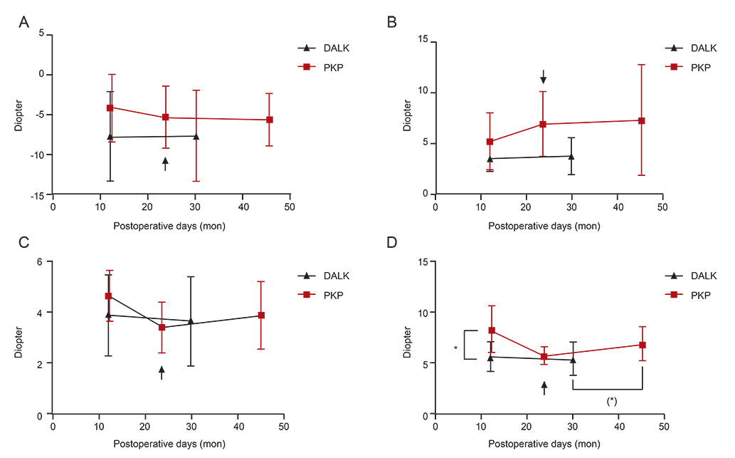
The follow-up period was 30 +/- 17 months in the DALK group and 45 +/- 20 months in the PKP group (p = 0.145). At final follow-up, the DALK and PKP groups achieved a BCVA (logarithm of the minimum angle of resolution) of 0.34 and 0.52, respectively (p = 0.980). Postoperative refractive error and mean simulated keratometric index showed myopic astigmatism in both groups without any statistical difference. Corneal irregularity index measured at 5 mm in the DALK group was less than that of the PKP group at 1-year follow-up (p = 0.021); however, at final follow-up, there was no longer a statistically significant difference. Endothelial cell counts were lower in the PKP group than in the DALK group at final follow-up (p = 0.021).
CONCLUSIONS
The optical outcomes of DALK with same-size grafts for keratoconus are comparable to those of PKP. Endothelial cell counts are more stable in DALK compared to PKP.
MeSH Terms
Figure
Cited by 1 articles
-
Long-term Results of Mini Asymmetric Radial Keratotomy and Corneal Cross-linking for the Treatment of Keratoconus
Marco Abbondanza, Gabriele Abbondanza, Valentina De Felice, Zoie Shui-Yee Wong
Korean J Ophthalmol. 2019;33(2):189-195. doi: 10.3341/kjo.2018.0028.
Reference
-
1. Pramanik S, Musch DC, Sutphin JE, Farjo AA. Extended long-term outcomes of penetrating keratoplasty for keratoconus. Ophthalmology. 2006; 113:1633–1638.2. Tay KH, Chan WK. Penetrating keratoplasty for keratoconus. Ann Acad Med Singapore. 1997; 26:132–137.3. Zadok D, Schwarts S, Marcovich A, et al. Penetrating keratoplasty for keratoconus: long-term results. Cornea. 2005; 24:959–961.4. Shimazaki J. The evolution of lamellar keratoplasty. Curr Opin Ophthalmol. 2000; 11:217–223.5. Terry MA. The evolution of lamellar grafting techniques over twenty-five years. Cornea. 2000; 19:611–616.6. Doyle SJ, Harper C, Marcyniuk B, Ridgway AE. Prediction of refractive outcome in penetrating keratoplasty for keratoconus. Cornea. 1996; 15:441–445.7. Perry HD, Foulks GN. Oversize donor buttons in corneal transplantation surgery for keratoconus. Ophthalmic Surg. 1987; 18:751–752.8. Wilson SE, Bourne WM. Effect of recipient-donor trephine size disparity on refractive error in keratoconus. Ophthalmology. 1989; 96:299–305.9. Shimmura S, Ando M, Ishioka M, et al. Same-size donor corneas for myopic keratoconus. Cornea. 2004; 23:345–349.10. Girard LJ, Esnaola N, Rao R, et al. Use of grafts smaller than the opening for keratoconic myopia and astigmatism: a prospective study. J Cataract Refract Surg. 1992; 18:380–384.11. Goble RR, Hardman Lea SJ, Falcon MG. The use of the same size host and donor trephine in penetrating keratoplasty for keratoconus. Eye (Lond). 1994; 8(Pt 3):311–314.12. Serdarevic ON, Renard GJ, Pouliquen Y. Penetrating keratoplasty for keratoconus: role of videokeratoscopy and trephine sizing. J Cataract Refract Surg. 1996; 22:1165–1174.13. Fontana L, Parente G, Tassinari G. Clinical outcomes after deep anterior lamellar keratoplasty using the big-bubble technique in patients with keratoconus. Am J Ophthalmol. 2007; 143:117–124.14. Funnell CL, Ball J, Noble BA. Comparative cohort study of the outcomes of deep lamellar keratoplasty and penetrating keratoplasty for keratoconus. Eye (Lond). 2006; 20:527–532.15. Noble BA, Agrawal A, Collins C, et al. Deep Anterior Lamellar Keratoplasty (DALK): visual outcome and complications for a heterogeneous group of corneal pathologies. Cornea. 2007; 26:59–64.16. Rabinowitz YS. Videokeratographic indices to aid in screening for keratoconus. J Refract Surg. 1995; 11:371–379.17. Lee MJ, Lee HJ, Wee WR, et al. Effects of intrastromal air injection compared to hydro-injection on keratocyte apoptosis. J Korean Ophthalmol Soc. 2007; 48:555–562.18. Anwar M, Teichmann KD. Big-bubble technique to bare Descemet's membrane in anterior lamellar keratoplasty. J Cataract Refract Surg. 2002; 28:398–403.19. Kubaloglu A, Sari ES, Unal M, et al. Long-term results of deep anterior lamellar keratoplasty for the treatment of keratoconus. Am J Ophthalmol. 2011; 151:760–767.e1.20. Fogla R, Padmanabhan P. Results of deep lamellar keratoplasty using the big-bubble technique in patients with keratoconus. Am J Ophthalmol. 2006; 141:254–259.21. Feizi S, Javadi MA, Jamali H, Mirbabaee F. Deep anterior lamellar keratoplasty in patients with keratoconus: big-bubble technique. Cornea. 2010; 29:177–182.22. Han DC, Mehta JS, Por YM, et al. Comparison of outcomes of lamellar keratoplasty and penetrating keratoplasty in keratoconus. Am J Ophthalmol. 2009; 148:744–751.e1.23. Bahar I, Kaiserman I, Srinivasan S, et al. Comparison of three different techniques of corneal transplantation for keratoconus. Am J Ophthalmol. 2008; 146:905–912.e1.24. Javadi MA, Feizi S, Yazdani S, Mirbabaee F. Deep anterior lamellar keratoplasty versus penetrating keratoplasty for keratoconus: a clinical trial. Cornea. 2010; 29:365–371.25. Cohen AW, Goins KM, Sutphin JE, et al. Penetrating keratoplasty versus deep anterior lamellar keratoplasty for the treatment of keratoconus. Int Ophthalmol. 2010; 30:675–681.26. Javadi MA, Feizi S, Rastegarpour A. Effect of vitreous length and trephine size disparity on post-DALK refractive status. Cornea. 2011; 30:419–423.27. Kim KH, Ahn K, Chung ES, Chung TY. Comparison of deep anterior lamellar keratoplasty and penetrating keratoplasty for keratoconus. J Korean Ophthalmol Soc. 2008; 49:222–229.28. Kim KH, Choi SH, Ahn K, et al. Comparison of refractive changes after deep anterior lamellar keratoplasty and penetrating keratoplasty for keratoconus. Jpn J Ophthalmol. 2011; 55:93–97.29. Ardjomand N, Hau S, McAlister JC, et al. Quality of vision and graft thickness in deep anterior lamellar and penetrating corneal allografts. Am J Ophthalmol. 2007; 143:228–235.30. Gere JM, Goodno BJ. Mechanics of materials. Mason: Cengage Learning;2011. p. 486–502.31. Watson SL, Ramsay A, Dart JK, et al. Comparison of deep lamellar keratoplasty and penetrating keratoplasty in patients with keratoconus. Ophthalmology. 2004; 111:1676–1682.
- Full Text Links
- Actions
-
Cited
- CITED
-
- Close
- Share
- Similar articles
-
- Clinical Evaluation of Full-thickness Deep Lamellar Keratoplasty
- Three Cases of Urrets-Zavalia Syndrome Following Deep Lamellar Keratoplasty (DLKP)
- Comparison of Deep Anterior Lamellar Keratoplasty and Penetrating Keratoplasty for Keratoconus
- A Case of Anterior Synechiolysis with Lamellar Corneal Dissection in Penetrating Keratoplasty
- Cataract Extraction after Penetrating Keratoplasty


