Emergence of virulent pseudorabies virus infection in Northern China
- Affiliations
-
- 1Laboratory of Porcine Viral Diseases, China National Research Center for Veterinary Medicine, Luoyang 471003, China. wurui1977@163.com
- 2Pulike Biological Engineering, Inc., Luoyang 471003, China.
- KMID: 1705568
- DOI: http://doi.org/10.4142/jvs.2013.14.3.363
Abstract
- Our investigation was conducted in order to verify a recent severe epidemic at several swine farms in northern China that indicated a newly emerging disease. Evidence confirmed that the epidemic was caused by a virulent Pseudorabies virus infection in swine herds.
Keyword
MeSH Terms
-
Animals
China/epidemiology
Enzyme-Linked Immunosorbent Assay/veterinary
Epidemics/*veterinary
Herpesvirus 1, Suid/classification/*isolation & purification/*pathogenicity
Pseudorabies/*epidemiology/mortality/pathology/virology
Reverse Transcriptase Polymerase Chain Reaction/veterinary
Sequence Analysis, DNA/veterinary
Swine
Swine Diseases/*epidemiology/mortality/pathology/virology
Vaccination/adverse effects/veterinary
Virulence
Figure
-

Fig. 1 Regions of pseudorabies virus (PRV) outbreak in China. The provinces or autonomous cities affected are indicated in pink. Regions marked with black triangles indicate areas showing positive PRV results as detected by enzyme-linked immunosorbent assay and polymerase chain reaction analyses.
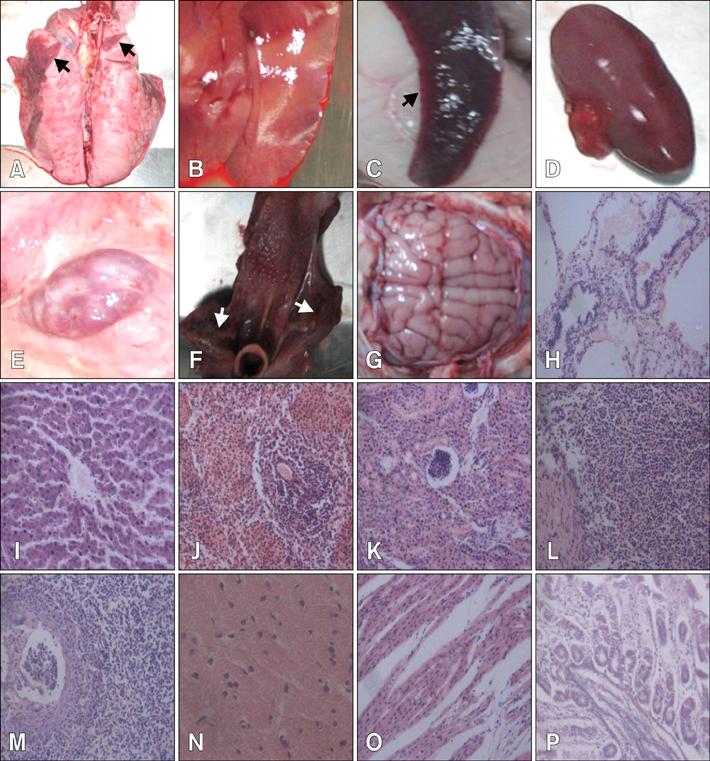
Fig. 2 Severe damage to multiple organs in experimental, infected piglets by postmortem and histopathological examinations. (A) Lung necrosis (arrows). (B) Liver with yellowish white spots indicating necrosis or hemorrhage. (C) Spleen infarct (arrow). (D) Kidney with bleeding spots. (E) Hemorrhagic lymph node. (F) Tonsil necrosis (arrows). (G) Slight encephalic edema. (H) Alveolar ducts and terminal bronchiolar cavities filled with cellular and serous exudates. (I) Swelling and degeneration of liver cells. (J) Splenic cord with unclear structure and reduced lymphocytes. (K) Swelling and disintegration of epithelial cells. (L) Reduced lymphoid nodules with irregular structures. (M) Epithelial cells filled with eosinophilic intranuclear inclusions. (N) Glial cells with neurons. (O) Breakage and disintegration of myocardial fibers. (P) Midgut gland atrophy. H&E stain, ×400.
Cited by 3 articles
-
Molecular characterization and phylogenetic analysis of pseudorabies virus variants isolated from Guangdong province of southern China during 2013–2014
Jindai Fan, Xiduo Zeng, Guanqun Zhang, Qiwen Wu, Jianqiang Niu, Baoli Sun, Qingmei Xie, Jingyun Ma
J Vet Sci. 2016;17(3):369-375. doi: 10.4142/jvs.2016.17.3.369.Seroprevalence and associated risk factors of pseudorabies in Shandong province of China
Dongfang Hu, Lin Lv, Zhendong Zhang, Yihong Xiao, Sidang Liu
J Vet Sci. 2016;17(3):361-368. doi: 10.4142/jvs.2016.17.3.361.Functional analysis of prv-miR-LLT11a encoded by pseudorabies virus
Huimin Liu, Li Yang, Zhibin Shi, Ruiqi Lv, Xia Yang, Chuanqing Wang, Lu Chen, Hongtao Chang
J Vet Sci. 2019;20(6):. doi: 10.4142/jvs.2019.20.e68.
Reference
-
1. Boadella M, Gortázar C, Vicente J, Ruiz-Fons F. Wild boar: an increasing concern for Aujeszky's disease control in pigs. BMC Vet Res. 2012; 8:7.
Article2. Cramer SD, Campbell GA, Njaa BL, Morgan SE, Smith SK 2nd, McLin WR 4rd, Brodersen BW, Wise AG, Scherba G, Langohr IM, Maes RK. Pseudorabies virus infection in Oklahoma hunting dogs. J Vet Diagn Invest. 2011; 23:915–923.
Article3. Marcaccini A, Peña ML, Quiroga MI, Bermúdez R, Nieto JM, Alemañ N. Pseudorabies virus infection in mink: a host-specific pathogenesis. Vet Immunol Immunopathol. 2008; 124:264–273.
Article4. Müller T, Hahn EC, Tottewitz F, Kramer M, Klupp BG, Mettenleiter TC, Freuling C. Pseudorabies virus in wild swine: a global perspective. Arch Virol. 2011; 156:1691–1705.
Article
- Full Text Links
-
- Actions
-
Cited
- CITED
-
- Close
- Share
-
- Similar articles
-
- Seroprevalence and associated risk factors of pseudorabies in Shandong province of China
- Functional analysis of prv-miR-LLT11a encoded by pseudorabies virus
- Molecular characterization and phylogenetic analysis of pseudorabies virus variants isolated from Guangdong province of southern China during 2013–2014
- Epidemiological investigation of porcine pseudorabies virus and its coinfection rate in Shandong Province in China from 2015 to 2018
- Comparative proteomic analysis of Virology PK-15 cells infected with wild-type strain and its EP0 gene-deleted mutant strain of pseudorabies virus
