Clin Orthop Surg.
2013 Jun;5(2):152-154. 10.4055/cios.2013.5.2.152.
Symptomatic Os Infranaviculare
- Affiliations
-
- 1Department of Orthopedic Surgery, Ewha Womans University School of Medicine, Seoul, Korea. kimjk@ewha.ac.kr
- KMID: 1705538
- DOI: http://doi.org/10.4055/cios.2013.5.2.152
Abstract
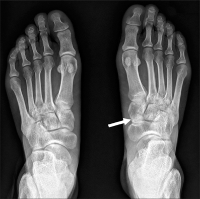
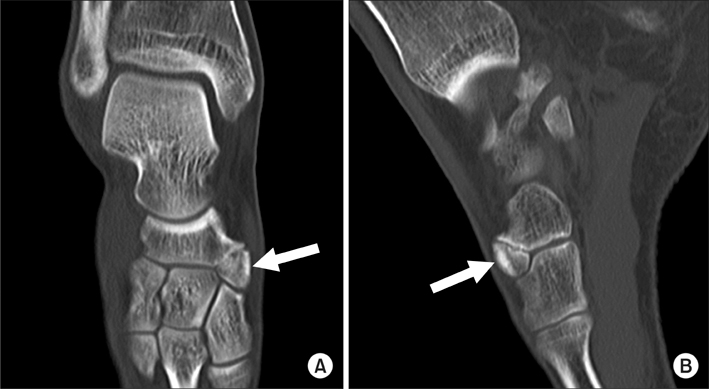
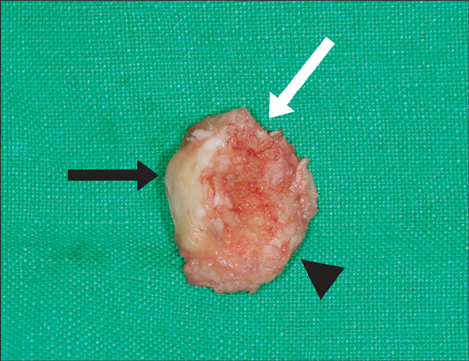
- The author observed a new accessory bone of the foot in the distal portion of navicular, which articulated with the medial cuneiform and the intermediate cuneiform, and named it os infranaviculare. A degenerative change was observed between the accessory bone and the navicular; this caused midfoot pain to the patient during weight-bearing. Thus, the patient was treated by excision of the accessory bone. The symptom was relieved at one-year postoperative.
Keyword
MeSH Terms
Figure
Cited by 1 articles
-
Symptomatic Os Paracuneiforme: A Case Report
Seung Hun Woo, Won Chul Shin
J Korean Foot Ankle Soc. 2021;25(2):108-110. doi: 10.14193/jkfas.2021.25.2.108.
Reference
-
1. Coskun N, Yuksel M, Cevener M, et al. Incidence of accessory ossicles and sesamoid bones in the feet: a radiographic study of the Turkish subjects. Surg Radiol Anat. 2009. 31(1):19–24.
Article2. Mellado JM, Ramos A, Salvadó E, Camins A, Danus M, Sauri A. Accessory ossicles and sesamoid bones of the ankle and foot: imaging findings, clinical significance and differential diagnosis. Eur Radiol. 2003. 13:Suppl 6. L164–L177.
Article3. Tsuruta T, Shiokawa Y, Kato A, et al. Radiological study of the accessory skeletal elements in the foot and ankle. Nihon Seikeigeka Gakkai Zasshi. 1981. 55(4):357–370.4. de Clercq PF, Bevernage BD, Leemrijse T. Stress fracture of the navicular bone. Acta Orthop Belg. 2008. 74(6):725–734.5. DiGiovanni CW, Patel A, Calfee R, Nickisch F. Osteonecrosis in the foot. J Am Acad Orthop Surg. 2007. 15(4):208–217.
Article6. Mair SD, Coogan AC, Speer KP, Hall RL. Gout as a source of sesamoid pain. Foot Ankle Int. 1995. 16(10):613–616.
Article7. Prescher A. Some remarks on, and a new case of the rare os intercuneiforme (Dwight). Ann Anat. 1997. 179(4):317–320.
Article8. Miller TT. Painful accessory bones of the foot. Semin Musculoskelet Radiol. 2002. 6(2):153–161.
Article


