J Clin Neurol.
2006 Jun;2(2):141-145. 10.3988/jcn.2006.2.2.141.
Diffusion-Weighted MRI in Recurrent Wernicke's Encephalopathy: a Remarkable Cerebellar Lesion
- Affiliations
-
- 1Department of Neurology, Konyang University College of Medicine, Daejeon, Korea. jekim220@medimail.co.kr
- 2Department of Neurology, Eulji University College of Medicine, Daejeon, Korea.
- 3Department of Radiology, Eulji University College of Medicine, Daejeon, Korea.
- KMID: 1700748
- DOI: http://doi.org/10.3988/jcn.2006.2.2.141
Abstract
- We report unusual MRI findings (including those from diffusion-weighted imaging (DWI)) in a patient with recurrent Wernicke's encephalopathy with a remarkable cerebellar lesion. DWI showed high signal intensities in the superior portion of the cerebellar hemisphere and vermis area. After thiamine administration, clinical symptoms improved and the lesions with high signal intensities disappeared on follow-up DWI.
Keyword
MeSH Terms
Figure
-
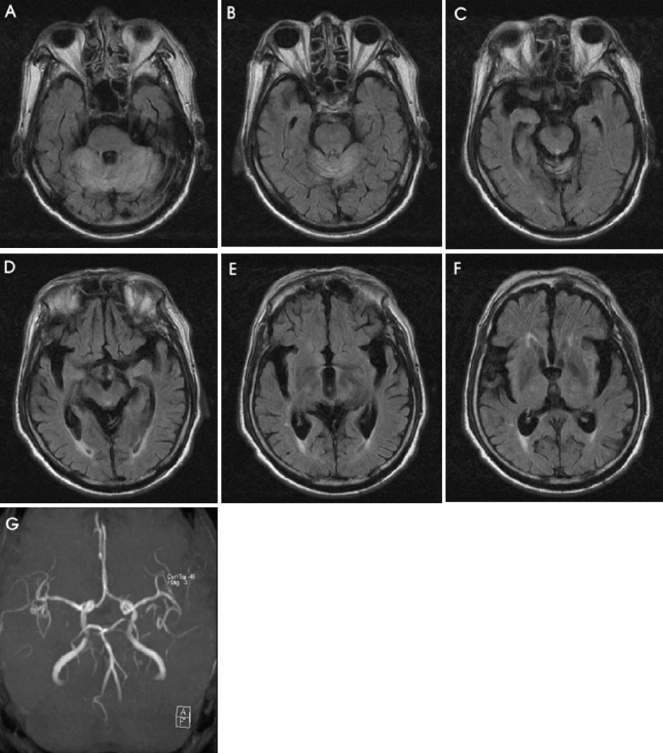
Figure 1 Axial FLAIR images (TR=6500 ms, TE=105 ms) show bilateral symmetrically distributed hyperintensity in the cerebellar hemisphere (A) and the vermis (B, C), and severe atrophy of mammillary body, periaqueduct, and medial thalamus (D, E, F). (G) Bilateral superior cerebellar arteries seem to be patent on MR angiography.
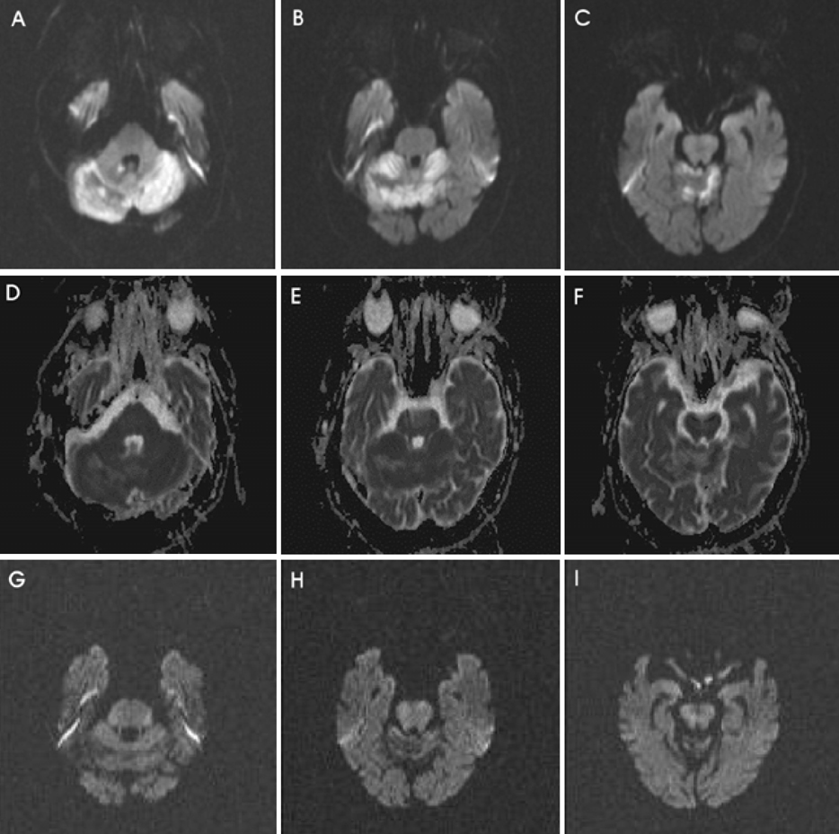
Figure 2 DWI images (A, B, C) show bright, high signal intensities in the bilateral cerebellum, while the corresponding apparent diffusion-coefficient values of the lesions (D, E, F) show low signal intensities. Follow-up DWI after 2 weeks of thiamine shows complete recovery of cerebellar lesion (G, H, I).
Reference
-
1. Reuler JB, Girard DE, Cooney TG. Current concepts. Wernicke's encephalopathy. N Engl J Med. 1985. 312:1035–1039.2. Victor M, Adams RD, Collins GH. The Wernicke-Korsakoff syndrome and related neurologic disorders due to alcoholism and malnutrition. 12 Contemporary Neurology Series. 1989. 2nd edn. Philadelphia: FA Davis.3. Murata T, Fujito T, Kimura H, Omori M, Itoh H, Wada Y. Serial MRI and (1)H-MRS of Wernicke's encephalopathy: report of a case with remarkable cerebellar lesions on MRI. Psychiatry Res. 2001. 108:49–55.
Article4. Doherty MJ, Watson NF, Uchino K, Hallam DK, Cramer SC. Diffusion abnormalities in patients with Wernicke encephalopathy. Neurology. 2002. 58:655–657.
Article5. Bergui M, Bradac GB, Zhong JJ, Barbero PA, Durelli L. Diffusion-weighted MR in reversible Wernicke encephalopathy. Neuroradiology. 2001. 43:969–972.
Article6. Chu K, Kang DW, Kim HJ, Lee YS, Park SH. Diffusionweighted imaging abnormalities in Wernicke encephalopathy: reversible cytotoxic edema? Arch Neurol. 2002. 59:123–127.
Article7. Niclot P, Guichard JP, Djomby R, Sellier P, Bousser MG, Chabriat H. Transient decrease of water diffusion in Wernicke's encephalopathy. Neuroradiology. 2002. 44:305–307.
Article8. Watson WD, Verma A, Lenart MJ, Quast TM, Gauerke SJ, McKenna GJ. MRI in acute Wernicke's encephalopathy. Neurology. 2003. 61:527.
Article9. Opdenakker G, Gelin G, De Surgeloose D, Palmers Y. Wernicke encephalopathy: MR findings in two patients. Eur Radiol. 1999. 9:1620–1624.
Article10. Suzuki S, Ichijo M, Fujii H, Matsuoka Y, Ogawa Y. Acute Wernicke's encephalopathy: comparison of magnetic resonance images and autopsy findings. Intern Med. 1996. 35:831–834.
Article11. Dreyfus DM. Thiamine and the nervous system: an overview. J Nutr Sci Vitaminol (Tokyo). 1976. 22:Suppl. 13–16.12. Witt ED. Neuroanatomical consequences of thiamine deficiency: a comparative analysis. Alcohol Alcohol. 1985. 20:201–221.13. Butterworth RF, Kril JJ, Harper CG. Thiamine-dependent enzyme changes in the brains of alcoholics: relationship to the Wernicke-Korsakoff syndrome. Alcohol Clin Exp Res. 1993. 17:1084–1088.
Article14. Schaefer PW, Grant PE, Gonzalez RG. Diffusion-weighted MR imaging of the brain. Radiology. 2000. 217:331–345.
Article
- Full Text Links
-
- Actions
-
Cited
- CITED
-
- Close
- Share
-
- Similar articles
-
- Diffusion Weighted Magnetic Resonance Imaging in a Patient with Acute Wernicke Encephalopathy
- Wernicke Encephalopathy Associated with Acute Wet Beriberi
- A Case of Wernicke's Encephalopathy in a Patient with Multiple System Atrophy
- Two Cases of Wernicke's Encephalopathy with Hyperemesis Gravidarum
- Diffusion-Weighted MR Imaging in Acute Wernicke's Encephalopathy Associated with Pseudomembranous Colitis: A Case Report and Review of the Literature
