Korean J Gastroenterol.
2013 Mar;61(3):160-165. 10.4166/kjg.2013.61.3.160.
Acute Extensive Ischemic Enteritis in a Young Man Diagnosed with Wireless Capsule Endoscopy: A Case Report
- Affiliations
-
- 1Department of Internal Medicine, Jeju National University School of Medicine, Jeju, Korea. songhj@jejunu.ac.kr
- 2Department of Radiology, Jeju National University School of Medicine, Jeju, Korea.
- KMID: 1501639
- DOI: http://doi.org/10.4166/kjg.2013.61.3.160
Abstract
- Ischemic enteritis is caused by either the interruption or significant reduction of arterial inflow to the small intestine. Risk factors are old age, diabetes mellitus and cardiovascular disease. It is very rare in young patients. We experienced a 21-year-old man with recurrent acute ischemic enteritis who was diagnosed with capsule endoscopy. He had previously taken medications for pulmonary hypertension and obstruction of both carotid arteries, and about 20 months earlier, he had been admitted due to hematochezia. Two sessions of angiography did not reveal the cause of hematochezia. At that time, capsule endoscopy showed mucosal edema and erythema in the terminal ileum, suggesting healed ischemic enteritis. The patient was admitted again due to hematochezia. Abdominal computed tomography showed focal celiac trunk stenosis and diffuse wall thickening of the small intestine, suggesting ischemic enteritis. Capsule endoscopy showed multiple active ulcers and severe hemorrhage with exudate, extending from the proximal jejunum to the terminal ileum. Using capsule endoscopy, the patient was diagnosed with acute extensive ischemic enteritis. Because endoscopic images of ischemic enteritis have rarely been reported, we report a case of a 21-year-old man who was diagnosed acute extensive ischemic enteritis with capsule endoscopy.
Keyword
MeSH Terms
Figure
-
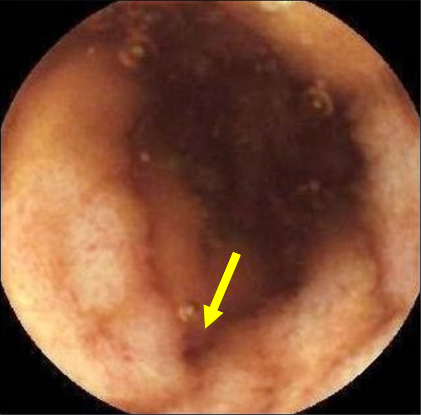
Fig. 1. Capsule endoscopic findings obtained 20 months earlier before admission. Mild mucosal edema with hyperemia in the terminal ileum (yellow arrow) suggested ischemic enteritis in the recovery phase.
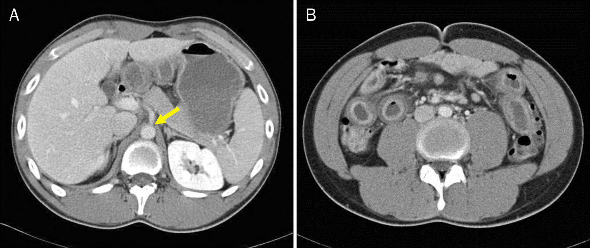
Fig. 2. Abdominal pelvic CT showing celiac trunk stenosis (A) (yellow arrow), diffuse wall thickening of the small bowel (B), which were suggestive of extensive ischemic enteritis.
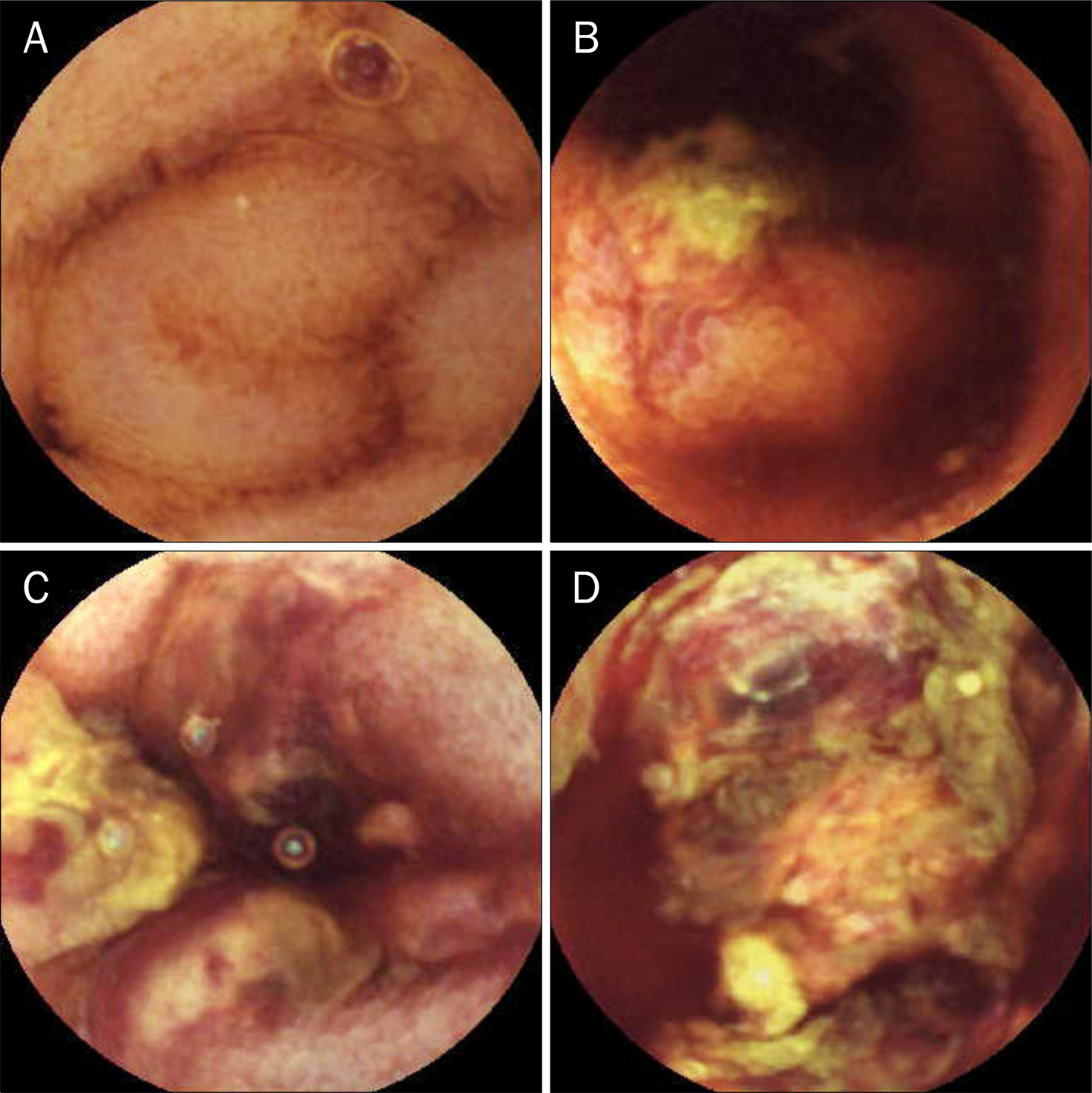
Fig. 3. Capsule endoscopy findings on admission. (A) Diffusely swollen mucosa was seen in the duodenum. (B) Active ulcers with exudates were noted in the proximal jejunum. (C, D) Severe mucosal hemorrhage with desquamation was seen, extending from the proximal jejunum to the terminal ileum.
Reference
-
References
1. Delikoukos S, Christodoulidis G, Zacharoulis D, Poultsidi A, Hatzitheofilou C. Multiple small bowel ruptures due to ischemic enteritis: a case report. World J Gastroenterol. 2006; 12:4262–4263.
Article2. Díaz Nieto R, Varcada M, Ogunbiyi OA, Winslet MC. Systematic review on the treatment of ischaemic colitis. Colorectal Dis. 2011; 13:744–747.
Article3. Nishimura N, Yamamoto H, Yano T, et al. Balloon dilation when using double-balloon enteroscopy for smallbowel strictures associated with ischemic enteritis. Gastrointest Endosc. 2011; 74:1157–1161.
Article4. Clavien PA. Diagnosis and management of mesenteric infarction. Br J Surg. 1990; 77:601–603.
Article5. Renner P, Kienle K, Dahlke MH, et al. Intestinal ischemia: current treatment concepts. Langenbecks Arch Surg. 2011; 396:3–11.
Article6. Yang XY, Chen CX, Zhang BL, et al. Diagnostic effect of capsule endoscopy in 31 cases of subacute small bowel obstruction. World J Gastroenterol. 2009; 15:2401–2405.
Article7. Liatsos C, Goulas S, Karagiannis S, Patelaros E, Sabaziotis D, Mavrogiannis C. Diagnosis of smallbowel ischemic necrosis by capsule endoscopy. Gastrointest Endosc. 2005; 62:439–440.
Article8. Levy PJ, Krausz MM, Manny J. Acute mesenteric ischemia: improved results–a retrospective analysis of ninety-two patients. Surgery. 1990; 107:372–380.9. Kitchens CS. Evolution of our understanding of the pathophysiology of primary mesenteric venous thrombosis. Am J Surg. 1992; 163:346–348.
Article10. Eryigit E, Hoentjen F, Barbe E, van Meyel JJ. Intestinal ischaemia caused by mesenteric inflammatory veno-occlusive disease. Neth J Med. 2008; 66:486–488.11. Brandt LJ, Boley SJ. AGA technical review on intestinal ischemia. American Gastrointestinal Association. Gastroenterology. 2000; 118:954–968.12. Aschoff AJ, Stuber G, Becker BW, et al. Evaluation of acute mesenteric ischemia: accuracy of biphasic mesenteric multidetector CT angiography. Abdom Imaging. 2009; 34:345–357.
Article13. Mönkemüller K, Bellutti M, Malfertheiner P. Small-bowel endoscopy. Endoscopy. 2007; 39:978–985.
Article14. Pennazio M, Santucci R, Rondonotti E, et al. Outcome of patients with obscure gastrointestinal bleeding after capsule endoscopy: report of 100 consecutive cases. Gastroenterology. 2004; 126:643–653.
Article15. Rondonotti E, Villa F, Mulder CJ, Jacobs MA, de Franchis R. Small bowel capsule endoscopy in 2007: indications, risks and limitations. World J Gastroenterol. 2007; 13:6140–6149.
Article16. Chan FS, Chu KM. Capsule endoscopy for gastrointestinal bleeding of obscure origin. Asian J Surg. 2008; 31:96–99.
Article17. Hashimoto Y, Endo Y, Kuroki Y, Yoshikumi H, Yoshiba M. Transient ischemic smallbowel ulcers secondary to acute superior mesenteric artery branch thromboembolism diagnosed by double balloon enteroscopy. Endoscopy. 2008; 40(Suppl 2):E161.
Article18. Lee KH, Park JM, Chun SW, et al. A case of acute ischemic enteritis caused by polyarteritis nodosa. Korean J Gastrointest Endosc. 2006; 33:303–306.19. Park JH, Jung YS, Kim YK, et al. A case of Churg-Strauss syndrome with interstinal perforation. Tuberc Respir Dis. 2009; 66:374–379.
Article20. Lin HP, Wang YM, Huo AP. Severe, recurrent lupus enteritis as the initial and only presentation of systemic lupus erythematosus in a middle-aged woman. J Microbiol Immunol Infect. 2011; 44:152–155.
Article
- Full Text Links
-
- Actions
-
Cited
- CITED
-
- Close
- Share
-
- Similar articles
-
- A Case of Acute Small Bowel Bleeding Diagnosed by Capsule Endoscopy
- A Case of Small Bowel Polyp Bleeding Diagnosed by Capsule Endoscopy
- Capsule Endoscopy in Children
- A Case of Bleeding Meckel's Diverticulm Diagnosed by Wireless Capsule Endoscopy
- Retrieval of a Retained Capsule due to Isolated Crohn's Enteritis by Means of Double Balloon Enteroscopy
