Astigmatic Changes after Horizontal Rectus Muscle Surgery in Intermittent Exotropia
- Affiliations
-
- 1Department of Ophthalmology & Visual Science, Bucheon St. Mary's Hospital, The Catholic University of Korea College of Medicine, Bucheon, Korea. nyeokang@catholic.ac.kr
- KMID: 1499682
- DOI: http://doi.org/10.3341/kjo.2012.26.6.438
Abstract
- PURPOSE
To evaluate the changes of refractive astigmatism after horizontal rectus muscle surgery in intermittent exotropic children.
METHODS
Sixty-nine exotropic patients were retrospectively reviewed. Of those, 35 patients received unilateral lateral rectus recession (BLR group, 35 eyes) and 34 patients received unilateral lateral rectus recession and medial rectus resection (R&R group, 34 eyes). Non-cycloplegic refractions were measured until 6 months postoperatively. Spherical equivalent (SE), J0 and J45 using power vectors were calculated to determine and compare the changes of refractive astigmatism and axis in both groups.
RESULTS
SE significantly decreased after surgery for the first week and did not changed thereafter in both groups (p = 0.000 and p = 0.018, respectively). In BLR group, J0 showed significant changes at the first week and 1 month after surgery (p = 0.005 and p = 0.016, respectively), but in R&R group, J0 changed significantly between 1 week and 3 months postoperatively (p = 0.023 and p = 0.016, respectively). J45 did not change significantly as time passed in both groups (all p > 0.05). There was no statistically significant difference in the magnitude of changes in SE, J0 and J45 between the two groups after the 6-month follow-up (p = 0.500, p = 0.244 and p = 0.202, respectively).
CONCLUSIONS
Horizontal rectus muscle surgery in intermittent exotropic children tends to induce a statistically significant change in astigmatism in the with-the-rule direction and myopic shift in SE. This astigmatism change seems to occur within the first 3 months after surgery. Thus, astigmatism induced by surgery should be checked and corrected at least 3 months after horizontal strabismus surgery.
MeSH Terms
Figure
-

Fig. 1 Changes of spherical equivalent (SE), J0, and J45 after surgery according to the postoperative time course in bilateral lateral rectus recession (BLR) group (A,C,E) and unilateral lateral rectus recession and medial rectus resection (R&R) group (B,D,F). SE significantly decreased at 1 week postoperatively (p = 0.000 and p = 0.018, respectively) and did not change significantly thereafter in two groups (A,B). In BLR group, J0 showed significant changes until the first month after surgery (p = 0.005 and p = 0.016, respectively) (C). In R&R group, J0 showed significant changes between 1 week postoperatively and 3 months postoperatively (p = 0.023 and p = 0.016 respectively) (D). J45 did not show any significant changes (p > 0.05) in both groups (E,F). POD= postoperative day.
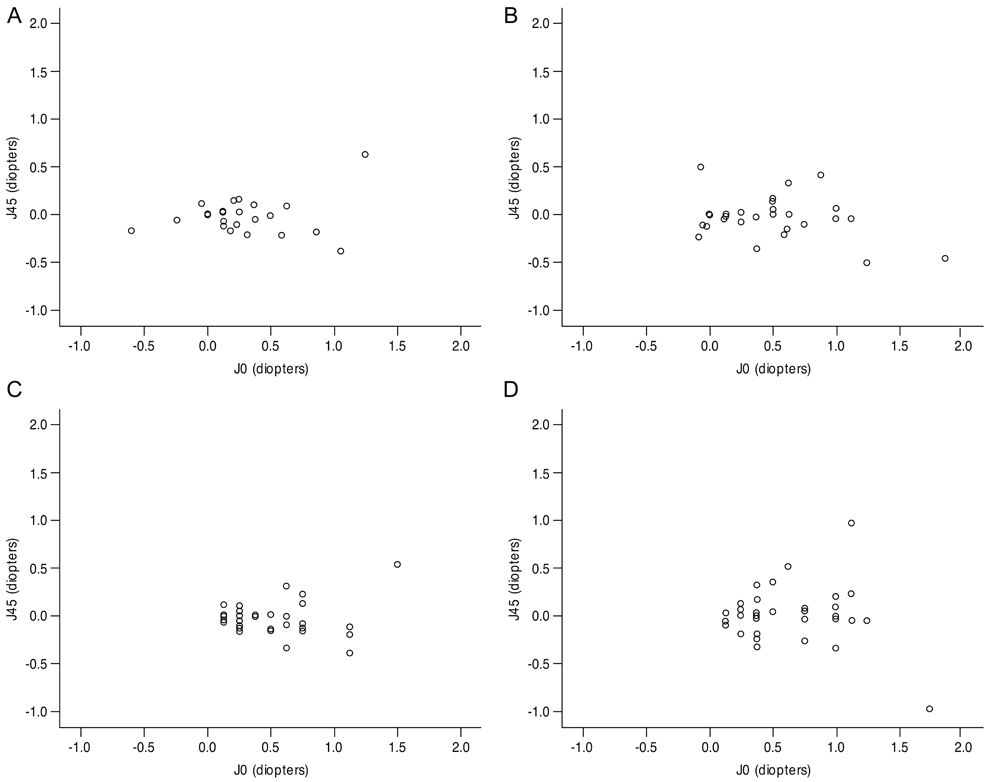
Fig. 2 Distribution of J0's and J45's before surgery (A,B) and at 6 months postoperatively (C,D) in bilateral lateral rectus recession group (A,C) and unilateral lateral rectus recession and medial rectus resection group (B,D). Non-cycloplegic astigmatism before surgery increased on average after surgery in both groups. Each data point represents the astigmatism component of power vector.
Reference
-
1. Denis D, Bardot J, Volot F, et al. Effects of strabismus surgery on refraction in children. Ophthalmologica. 1995. 209:136–140.2. Kwitko S, Feldon S, McDonnell PJ. Corneal topographic changes following strabismus surgery in Grave's disease. Cornea. 1992. 11:36–40.3. Snir M, Nissenkorn I, Buckman G, et al. Postoperative refractive changes in children with congenital esotropia: a preliminary study. Ophthalmic Surg. 1989. 20:57–62.4. Preslan MW, Cioffi G, Min YI. Refractive error changes following strabismus surgery. J Pediatr Ophthalmol Strabismus. 1992. 29:300–304.5. Killer HE, Bahler A. Significant immediate and long-term reduction of astigmatism after lateral rectus recession in divergent Duane's syndrome. Ophthalmologica. 1999. 213:209–210.6. Reynolds RD, Nelson LB, Greenwald M. Large refractive change after strabismus surgery. Am J Ophthalmol. 1991. 111:371–372.7. Nardi M, Rizzo S, Pellegrini G, Lepri A. Effects of strabismus surgery on corneal topography. J Pediatr Ophthalmol Strabismus. 1997. 34:244–246.8. Marshall D. Changes in refraction following operation for strabismus. Arch Ophthalmol. 1936. 15:1020–1031.9. Fix A, Baker JD. Refractive changes following strabismus surgery. Am Orthopt J. 1985. 35:59–62.10. Kwito S, Sawusch MR, McDonnell PJ, et al. Effect of extraocular muscle surgery on corneal topography. Arch Ophthalmol. 1991. 109:873–878.11. Rajavi Z, Mohammad Rabei H, Ramezani A, et al. Refractive effect of the horizontal rectus muscle recession. Int Ophthalmol. 2008. 28:83–88.12. Thibos LN, Wheeler W, Horner D. Power vectors: an application of Fourier analysis to the description and statistical analysis of refractive error. Optom Vis Sci. 1997. 74:367–375.13. Thibos LN, Horner D. Power vector analysis of the optical outcome of refractive surgery. J Cataract Refract Surg. 2001. 27:80–85.14. Yoo JM, Ryoo MH. The changes of astigmatism following horizontal strabismus surgery. J Korean Ophthalmol Soc. 1990. 31:337–341.15. Lee JH, Choi DG. The changes of refractive error and corneal astigmatism after horizontal strabismus surgery. J Korean Ophthalmol Soc. 1995. 36:1221–1227.16. Hainsworth DP, Bierly JR, Schmeisser ET, Baker RS. Corneal topographic changes after extraocular muscle surgery. J AAPOS. 1999. 3:80–86.17. Paik HJ, Park SO. Corneal topographic changes after horizontal rectus muscles surgery. J Korean Ophthalmol Soc. 1999. 40:3187–3194.18. Mun GH, Heo H, Park SW, Park YG. The changes of corneal astigmatism and refraction after horizontal rectus muscle surgery in intermittent exotropia. J Korean Ophthalmol Soc. 2010. 51:581–587.19. Thompson WE, Reinecke RD. The changes in refractive status following routine strabismus surgery. J Pediatr Ophthalmol Strabismus. 1980. 17:372–374.20. Lee YC, Yoon JW, Lee WY. Refractive changes following strabismus surgery. J Korean Ophthalmol Soc. 1994. 35:704–708.21. Lee SY, Jeon SJ, Kim KS. Changes in refractive error following strabismus surgery. J Korean Ophthalmol Soc. 1997. 38:1262–1267.22. Bagheri A, Farahi A, Guyton DL. Astigmatism induced by simultaneous recession of both horizontal rectus muscles. J AAPOS. 2003. 7:42–46.23. Dottan SA, Hoffman P, Oliver MD. Astigmatism after strabismus surgery. Ophthalmic Surg. 1988. 19:128–129.24. Gordon RA, Donzis PB. Refractive development of the human eye. Arch Ophthalmol. 1985. 103:785–789.25. Dobson V, Fulton AB, Sebris SL. Cycloplegic refractions of infants and young children: the axis of astigmatism. Invest Ophthalmol Vis Sci. 1984. 25:83–87.26. Lieberman DM, Grierson JW. The lids influence on corneal shape. Cornea. 2000. 19:336–342.27. Read SA, Collins MJ, Carney LG. The influence of eyelid morphology on normal corneal shape. Invest Ophthalmol Vis Sci. 2007. 48:112–119.28. Zhao J, Mao J, Luo R, et al. Accuracy of noncycloplegic autorefraction in school-age children in China. Optom Vis Sci. 2004. 81:49–55.
- Full Text Links
-
- Actions
-
Cited
- CITED
-
- Close
- Share
-
- Similar articles
-
- Treatment of Sub-V Pattern Intermittent Exotropia
- The Changes of Corneal Astigmatism and Refraction After Horizontal Rectus Muscle Surgery in Intermittent Exotropia
- Postoperative Exotropic Drift: Comparison of Surgical Methods Combined with Lateral Rectus Muscle Recession in Exotropia
- The Clinical Analysis of Surgical Methods in Intermittent Exotropia
- The Effect of Unilateral Medial Rectus Muscle Resection in Patients with Recurrent Exotropia
