Endocrinol Metab.
2012 Jun;27(2):119-120. 10.3803/EnM.2012.27.2.119.
A Case of Asymptomatic Giant Cystic Pheochromocytoma
- Affiliations
-
- 1Department of Internal Medicine, Kangwon National University School of Medicine, Chuncheon, Korea. ehcho@kangwon.ac.kr
- 2Department of Anatomic Pathology, Kangwon National University School of Medicine, Chuncheon, Korea.
- KMID: 1497649
- DOI: http://doi.org/10.3803/EnM.2012.27.2.119
Abstract
- No abstract available.
MeSH Terms
Figure
-
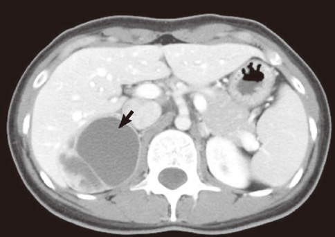
Fig. 1 Contrast-enhanced computed tomography scan shows about 6.7 cm sized round low density lesion with internal enhancing septum and a tiny calcification in the right suprarenal region (arrow).
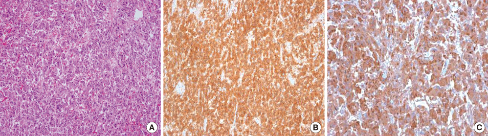
Fig. 2 A. Microscopic findings of tumor cells (H&E stain, × 200). B, C. Immunohistochemistry stain of pheochromocytoma (× 200). The tumor cells are strongly positive for synaptophysin (B) and chromogranin (C).
Reference
-
1. Erickson LA, Lloyd RV, Hartman R, Thompson G. Cystic adrenal neoplasms. Cancer. 2004. 101:1537–1544.2. Foster DG. Adrenal cysts. Review of literature and report of case. Arch Surg. 1966. 92:131–143.3. Scheible W, Coel M, Siemers PT, Siegel H. Percutaneous aspiration of adrenal cysts. AJR Am J Roentgenol. 1977. 128:1013–1016.4. Esquivel E Jr, Grabstald H. Giant adrenal cyst. J Urol. 1965. 94:635–638.5. Oh MM, Jung CS, Myoung SC, Moon WC. A case of cystic pheochromocytoma. Korean J Urol. 1990. 31:772–776.
