Bilateral Congenital Pulmonary Vein Stenosis with a Normal Connection
- Affiliations
-
- 1Department of Pediatrics, Chonnam National University Hospital, Chonnam National University Research Institute of Medical Sciences, Gwangju, Korea. cardiol@jnu.ac.kr
- 2Department of Pediatrics, Kwangju Christian Hospital, Gwangju, Korea.
- 3Department of Pediatrics, Seoul National University Children's Hospital, Seoul, Korea.
- KMID: 1491042
- DOI: http://doi.org/10.4250/jcu.2008.16.2.54
Abstract
- Congenital pulmonary vein stenosis (CPVS) with an anatomically normal connection is a rare cardiac malformation. This cardiac anomaly usually is accompanied by other cardiac abnormalities. Bilateral CPVS is a more severe form of a CPVS and it usually leads to progressive pulmonary hypertension and death if it is not treated. Here, we report a patient with a history of cough, tachypnea and hemoptysis and suspected CPVS due to an abnormal thoracic roentgenogram with dilated right pulmonary arteries and pulmonary cornus. The two-dimensional and color Doppler echocardiography demonstrated three stenosed pulmonary veins connected to the left atrium. However, the fourth vessel could not be visualized. There were no other cardiac malformations associated with the CPVS. The Technetium-99m macro-aggregate lung perfusion scan showed absent or diminished perfusion to the affected lobes of the lungs. In addition, the chest computed tomography with angiogram and cardiac catheterization confirmed the findings of the echocardiogram.
Keyword
MeSH Terms
Figure
-
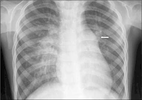
Fig. 1 The thoracic roentgenogram shows dilated right pulmonary arteries and pulmonary cornus (arrow) with relative radiolucency in the left lower lung field, reticular interstitial infiltrations in the right mid portion and a normal sized heart.
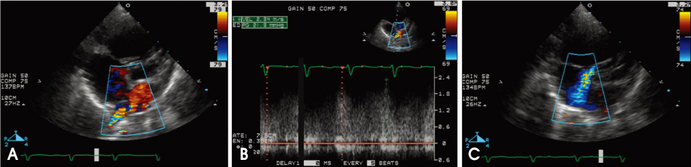
Fig. 2 The two-dimensional and color Doppler echocardiography. Three pulmonary veins appear connected to the left atrium with a continuous disturbed pulmonary venous flow pattern (A), the peak velocity was 2.3 m/sec without the normal phasic variation (B) as observed from the apical 4-chamber view. The right atrium was enlarged and there was no associated cardiac malformations except for a moderate tricuspid regurgitation (C).

Fig. 3 The Technetium-99m macro-aggregate lung perfusion scan shows absence of perfusion at the left lower lobe (black arrow) of the lung and diminished perfusion to the right middle lobe (white arrow) and lower lobe superior segment (arrow head) of the lung.

Fig. 4 The chest computed tomography with angiogram. The pulmonary vein draining from the right middle lobe (A) and right lower lobe (B) had focal stenosis (arrows). The one from the left upper lobe was draining with relatively normal flow but the one from the left lower lobe was not visible (C).
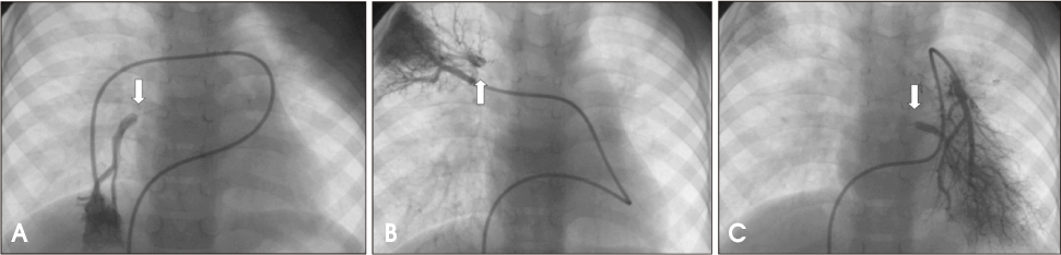
Fig. 5 The levophase of the pulmonary angiogram showed stenoses (arrows) of the right lower pulmonary vein (A), right upper pulmonary vein (B) and left lower pulmonary vein (C).
Reference
-
1. Anderson RH, Macartney FJ. Anderson RH, Baker EJ, Macartney FJ, Rigby ML, Shinebourne EA, Tynan MJ, editors. Pulmonary venous abnormalities. Pediatric Cardiology. 2001. 2nd Ed. London: Churchill Livingstone;890–894.2. Seale AN, Daubeney PE, Magee AG, Rigby ML. Pulmonary vein stenosis: initial experience with cutting balloon angioplasty. Heart. 2006. 92:815–820.
Article3. Bini RM, Cleveland DC, Ceballos R, Bargeron LM Jr, Pacifico AD, Kirklin JW. Congenital pulmonary vein stenosis. Am J Cardiol. 1984. 54:369–375.
Article4. Geva T, Van Praagh S. Allen HD, Driscoll DJ, Shaddy RE, Feltes TF, editors. Anomalies of the pulmonary veins. Moss and Adams' heart disease in infants, children, and adolescents: including the fetus and young adult. 2008. 7th Ed. Philadelphia: LWW;761–764.5. Mendeloff EN, Spray TL, Huddleston CB, Bridges ND, Canter CB, Mallory GB Jr. Lung transplantation for congenital pulmonary vein stenosis. Ann Thorac Surg. 1995. 60:903–906.
Article6. Jung CW, Lee SY, Yu J, Kim BJ, Yun TJ, Ko JK, Hong SJ. A Case of Congenital pulmonary vein stenosis diagnosed in an infant with recurrent hemoptysis. Pediatr Allergy Respir Dis (Korea). 2007. 17:434–439.7. Cho JJ, Park WS, Kang IS, Jun TG, Jung MJ. Congenital pulmonary vein stenosis manifested by severe cyanosis in infancy. Korean J Pediatr. 2004. 47:1114–1118.8. Lee JR, Lee JH, Kang CH, Noh CI, Seo JW. Unilateral pulmonary vein stenosis with life-threatening hemoptysis: a case report. Korean J Thorac Cardiovasc Surg. 2005. 38:725–728.9. Holcomb RG, Tyson RW, Ivy DD, Abman SH, Kinsella JP. Congenital pulmonary venous stenosis presenting as persistent pulmonary hypertension of the newborn. Pediatr Pulmonol. 1999. 28:301–306.
Article10. Adey CK, Soto B, Shin MS. Congenital pulmonary vein stenosis: a radiographic study. Radiology. 1986. 161:113–117.
Article11. Belcourt CL, Roy DL, Nanton MA, Finley JP, Gillis DA, Krause VW, Aterman K. Stenosis of individual pulmonary veins: radiologic findings. Radiology. 1986. 161:109–112.
Article12. Kingston HM, Patel RG, Watson GH. Unilateral absence or extreme hypoplasia of pulmonary veins. Br Heart J. 1983. 9:148–153.
Article13. Samdarshi TE, Morrow WR, Helmcke FR, Nanda NC, Bargeron LM Jr, Pacifico AD. Assessment of pulmonary vein stenosis by transesophagel echocardiography. Am Heart J. 1991. 122:1495–1498.
Article14. Chung NS, Ha JW, Yoon JH, Kim BO, Jang YS, Cho SY, Cho BK. Pulsed wave and color Doppler echocardiography and cardiac catheterization findings in bilateral pulmonary vein Stenosis. Korean Circ J. 1998. 28:647–652.
Article15. Bahl VK, Chandra S, Mishra S. Congenital stenosis of isolated pulmonary vein: role of retrograde pulmonary vein catheterization. Int J Cardiol. 1997. 60:103–105.
Article16. Bini RM, Bargeron LM Jr. Visualization of pulmonary vein obstruction by pulmonary artery wedge injection. Pediatr Cardiol. 1982. 2:161–162.
Article17. Driscoll DJ, Hesslein PS, Mullins CE. Congenital stenosis of individual pulmonary veins: clinical spectrum and unsuccessful treatment by transvenous balloon dilatation. Am J Cardiol. 1982. 49:1767–1772.
Article18. Kawashima Y, Ueda T, Naito Y, Morikawa E, Manabe H. Stenosis of pulmonary veins: report of a patient corrected surgically. Ann Thorac Surg. 1971. 12:196–202.19. Binet JP, Bouchard F, Langlois J, Chetochine F, Conso JF, Pottemain M. Unilateral congenital stenosis of the pulmonary veins: a very rare cause of pulmonary hypertension. J Thorac Cardiovasc Surg. 1972. 63:397–402.20. Mendelsohn AM, Bove EL, Lupinetti FM, Crowley DC, Lloyd TR, Fedderly RT, Beekman RH 3rd. Intraoperative and percutaneous stenting of congenital pulmonary artery and vein stenosis. Circulation. 1993. 88:II210–II217.21. Shin YH, Kim KE, Kwon HS, Yoo BW, Choi JY. Stent implantation to relieve secondary pulmonary venous stenosis in total anomalous pulmonary venous connection: case report. Korean J Pediatr. 2007. 50:919–924.
Article22. Devaney EJ, Chang AC, Ohye RG, Bove EL. Management of congenital and acquired pulmonary vein stenosis. Ann Thorac Surg. 2006. 81:992–995.
Article
- Full Text Links
-
- Actions
-
Cited
- CITED
-
- Close
- Share
-
- Similar articles
-
- Pulsed Wave and Color Doppler Echocardiography and Cardiac Catheterization Findings in Bilateral Pulmonary Vein Stenosis
- Congenital Pulmonary Vein Stenosis with Normal Anatomical Connection: One case report
- A Case of Surgically Corrected-Combined form of Total Anomalous Pulmonary Venous Return
- Congenital Pulmonary Vein Stenosis Manifested by Severe Cyanosis in Infancy
- Changes of Endothelin-1 after Pulmonary Venous Stenosis in Model
