Papillary Fibroelastoma of Pulmonary Valve Mimicking Infective Endocarditis
- Affiliations
-
- 1Department of Cardiology, Heart Center, Chonnam National University Hospital, Gwangju, Korea. jcpark54@hanmail.net
- KMID: 1486588
- DOI: http://doi.org/10.4250/jcu.2008.16.3.99
Abstract
- In this report, we describe a case of previous undiagnosed masses of the pulmonary valve mimicking infective endocarditis that were incidentally found during the work-up of a 62-year-old woman, who was presented with abdominal discomfort and dyspepsia. The pathologic findings were characteristics of a papillary fibroelastoma. Although benign, papillary fibroelastomas have the potential to cause lethal embolic events such as stroke, myocardial infarction, and pulmonary embolism are reported in some cases. Tumor identification and surgical excision are important to prevent such complications.
Keyword
MeSH Terms
Figure
-
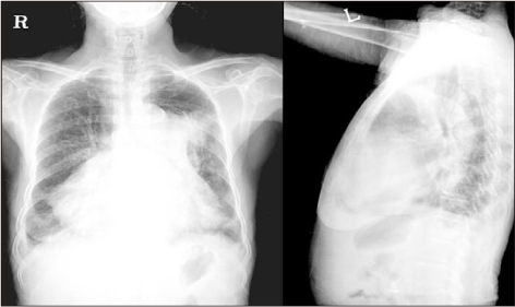
Fig. 1 Chest X-ray reveals marked cardiomegaly, especially right atrium, right ventricle with prominent pulmonary artery. Subsegmental atelectasis is noticed on left upper lung field.
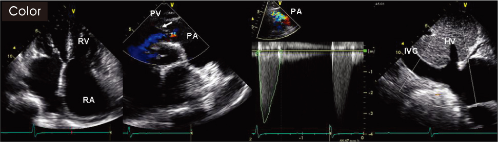
Fig. 2 Transthoracic echocardiography shows markedly dilated right atrium, right ventricle, pulmonary artery and inferior vena cava. Pulmonary valve stenosis and regurgitation are revealed (mild to moderate). PA: pulmonary artery, PV: pulmonary valve leaflet, IVC: inferior vena cava, HV: hepatic vein.
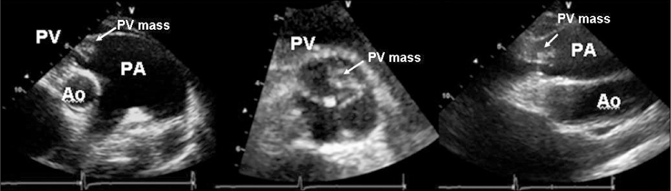
Fig. 3 Transthoracic echocardiography at the level of the pulmonary valve leaflet shows a 1.2×0.9 cm sized echogenic mass arising from the right cusp of the pulmonary valve. Ao: aorta, PA: pulmonary artery, PV: pulmonary valve leaflet.
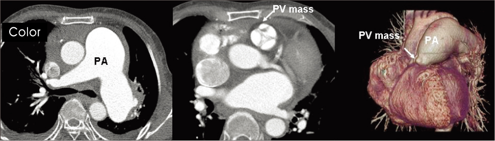
Fig. 4 Computer tomography shows dilated pulmonary artery and nodular mass on pulmonary valve. There is no filing defect suggesting pulmonary thrombiembolism.

Fig. 5 (A) Gross specimen shows multiple frondlike structures giving the appearance of a sea anemone. Histology of the lesion shows each frond core consists of a central fibroelastic stroma lined by flat endocardial cells. (B) Hematoxilin-eosin stain, original magnification, (C) type IV collagen stain, ×400, (D) masson-trichrom stain, original magnification.
Reference
-
1. Bevilacqua JA, Larrain E, Corredoira YA. Fibroelastoma of the mitral valve-a curable cause of stroke. Lancet Neurol. 2002. 1:389–390.
Article2. Howard RA, Aldea GS, Shapira OM, Kasznica JM, Davidoff R. Papillary fibroelastoma: increasing recognition of a surgical disease. Ann Thorac Surg. 1999. 68:1881–1885.
Article3. Lund GK, Schroder S, Koschyk DH, Nienaber CA. Echocardiographic diagnosis of papillary fibroelastoma of the mitral and tricuspid valve apparatus. Clin Cardiol. 1997. 20:175–177.
Article4. Burke A, Virmani R. Rosai J, editor. Papillary fibroelastoma. Atlas of tumor pathology, 3rd ed, fascicle 16, Tumors of the heart and great vessels. 1996. Washington, DC: Armed Forces Institute of Pathology;47–52.5. Boone SA, Campagna M, Walley VM. Lambl's excrescences and papillary fibroelastoma: are they different? Can J Cardiol. 1992. 8:372–376.6. Saad RS, Glavis CO, Bshara W, Liddicoat J, Dabbs DJ. Pulmonary valve papillary fibroelastoma. Arch Path Lab Meb. 2001. 124:933–934.
Article7. Grinda JM, Latremouille C, Berrebi A, Couetil JP, Chauvaud S, Fabiani JN, Deloche A, Carpentier A. Cardiac fibroelastoma. Six operated cases and review of the literature. Arch Mal Coeur Vaiss. 2000. 93:727–732.8. Kanarek SE, Wright P, Liu J, Boglioli LR, Bajwa AS, Hall M, Kort S. Multiple fibroelastomas: a case report and review of the literature. J Am Soc Echocardiogr. 2003. 16:373–376.
Article9. Weems WB, Aronson S, Yang X, Jayakar D, Jeevanandam V, Lang RM. Papillary fibroelastoma of the aortic valve. J Am Soc Echocardiogr. 2002. 15:382–384.
Article10. Rhee KS. A case of papillary fibroelastoma of the left ventricular outflow tract causing stroke. J Kor Soc Echo. 2004. 12:42–44.
Article11. Wantanabe T, Sasaki T, Kawamura H. Aortic valve papillary fibroelastoma; report of a case. Kyoby Geka. 2004. 57:226–228.12. Kanarek SE, Wright P, Liu J, Boglioli LR, Bajwa AS, Hall M, Kort S. Multiple fibroelastomas: a case report and review of the literature. J Am Soc Echocardiogr. 2003. 16:373–376.
Article13. Bhagwandien NS, Shah N, Costello JM, Gilbert CL, Blankenship JC. Echocardiographic detection of pulmonary valve papillary fibroelastoma. J Cardiovasc Surg (Torino). 1998. 39:351–354.14. Leonardi Cattolica FS, Minati A, Testa N, Sordini P, Costantino A, Gentili C, Alois A, Gallo R, Madaro P, Auriti A, Bernardi C, Staibano M. Two cardiac tumors with left ventricular location: myxoma and papillary fibroelastoma. Ital Heart J Suppl. 2004. 5:544–547.15. Gowda RM, Khan IA, Nair CK, Mehta NJ, Vasavada BC, Sacchi TJ. Cardiac papillary fibroelastoma: a comprehensive analysis of 725 cases. Am Heart J. 2003. 146:404–410.
Article
- Full Text Links
-
- Actions
-
Cited
- CITED
-
- Close
- Share
-
- Similar articles
-
- Tricuspid Papillary Fibroelastoma Mimicking Tricuspid Vegetation in a Patient with Severe Neutropenia
- Papillary Fibroelastoma Mimicking Vegetation of the Mitral Valve
- Aortic Valve Papillary Fibroelastoma Triggering Chest Pain: A case report
- Papillary Fibroelastoma of Pulmonary Valve with Congestive Heart Failure: A case report
- Infective Endocarditis with Patent Ductus Arteriosus at 60 Years Old Patient
