J Cardiovasc Ultrasound.
2012 Dec;20(4):213-215. 10.4250/jcu.2012.20.4.213.
Papillary Fibroelastoma Mimicking Vegetation of the Mitral Valve
- Affiliations
-
- 1Division of Cardiology, Department of Internal Medicine, Dong-A University College of Medicine, Busan, Korea. thpark65@dau.ac.kr
- 2Department of Pathology, Dong-A University College of Medicine, Busan, Korea.
- KMID: 2177412
- DOI: http://doi.org/10.4250/jcu.2012.20.4.213
Abstract
- Although cardiac papillary fibroelastoma is rare, it is the most common primary tumor of cardiac valves. The clinical presentation of these tumors varies from asymptomatic to embolic complications. We report an asymptomatic case of papillary fibroelastoma of mitral valve which was diagnosed by transthoracic echocardiography. The tumor was successfully resected by surgery.
MeSH Terms
Figure
-

Fig. 1 Transthoracic echocardiography shows a mobile and spherical mass of 1.0 cm in size attached to anterior mitral leaflet. Parasternal long axis view (A) and an apical four-chamberview (B).

Fig. 2 Gross specimen of excised mass reveals a friable mass with frond-like surface.
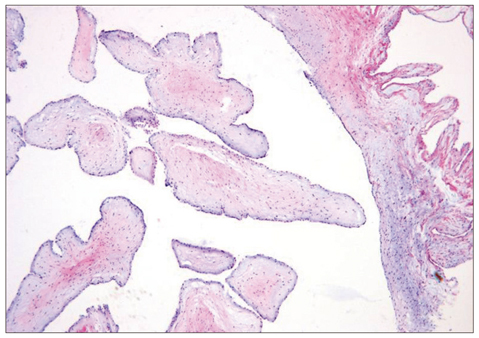
Fig. 3 Histological examination of the resected tumor showing papillary projection. The tumor surface is covered by a single layer of endocardial cells with an overlying thin layer of mucopolysaccharide matrix and an underlying acellular stroma composed of elastic fibers and collagen (Elastica van Gieson stain, × 40).
Reference
-
1. Reynen K. Frequency of primary tumors of the heart. Am J Cardiol. 1996. 77:107.
Article2. Mariscalco G, Bruno VD, Borsani P, Dominici C, Sala A. Papillary fibroelastoma: insight to a primary cardiac valve tumor. J Card Surg. 2010. 25:198–205.
Article3. Karaeren H, Ilgenli TF, Celik T, Deveci S, Kuralay E, Barçin C, Uzun M, Genç C, Demirtas E. Papillary fibroelastoma of the mitral valve with systemic embolization. Echocardiography. 2000. 17:165–167.
Article4. Liebeskind DS, Buljubasic N, Saver JL. Cardioembolic stroke due to papillary fibroelastoma. J Stroke Cerebrovasc Dis. 2001. 10:94–95.
Article5. Gowda RM, Khan IA, Nair CK, Mehta NJ, Vasavada BC, Sacchi TJ. Cardiac papillary fibroelastoma: a comprehensive analysis of 725 cases. Am Heart J. 2003. 146:404–410.
Article6. Grinda JM, Couetil JP, Chauvaud S, D'Attellis N, Berrebi A, Fabiani JN, Deloche A, Carpentier A. Cardiac valve papillary fibroelastoma: surgical excision for revealed or potential embolization. J Thorac Cardiovasc Surg. 1999. 117:106–110.
Article7. Boodhwani M, Veinot JP, Hendry PJ. Surgical approach to cardiac papillary fibroelastomas. Can J Cardiol. 2007. 23:301–302.
Article8. Gopaldas RR, Atluri PV, Blaustein AS, Bakaeen FG, Huh J, Chu D. Papillary fibroelastoma of the aortic valve: operative approaches upon incidental discovery. Tex Heart Inst J. 2009. 36:160–163.9. Sun JP, Asher CR, Yang XS, Cheng GG, Scalia GM, Massed AG, Griffin BP, Ratliff NB, Stewart WJ, Thomas JD. Clinical and echocardiographic characteristics of papillary fibroelastomas: a retrospective and prospective study in 162 patients. Circulation. 2001. 103:2687–2693.
Article10. Tanaka H, Narisawa T, Mori T, Masuda Y, Kishi D. Double primary left ventricular and aortic valve papillary fibroelastoma. Circ J. 2004. 68:504–506.
Article
- Full Text Links
-
- Actions
-
Cited
- CITED
-
- Close
- Share
-
- Similar articles
-
- Tricuspid Papillary Fibroelastoma Mimicking Tricuspid Vegetation in a Patient with Severe Neutropenia
- Anesthetic Management of the Excision of Left Ventricular Papillary Fibroelastoma : A case report
- Papillary Fibroelastoma Originating from the Left Ventricle: A case report
- A Remnant Mitral Subvalvular Apparatus Mimicking Aortic Valve Vegetation after Mitral Valve Replacement
- Aortic Valve Papillary Fibroelastoma Triggering Chest Pain: A case report
