A Case of Right Coronary Artery Hematoma Detected by Echocardiography after Percutaneous Coronary Intervention
- Affiliations
-
- 1Department of Medicine, Samsung Medical Center, Sungkyunkwan University School of Medicine, Seoul, Korea. parksmc@gmail.com
- KMID: 1486392
- DOI: http://doi.org/10.4250/jcu.2008.16.4.126
Abstract
- A 69-year-old man was admitted to undergo percutaneous coronary intervention (PCI) for chronic total occlusion of right coronary artery. He had diabetes mellitus, stable angina pectoris. Diagnostic coronary angiography demonstrated proximal total occlusion of right coronary artery. PCI was failed due to failure of balloon passage. Echocardiography was performed after PCI and thickened epicardial tissue at right atrioventricular groove was noted. It was highly echogenic and localized along the course of mid right coronary artery. In following echocardiogram after 12 days, the size of echogenic mass was decreased from 3.4 cmx2.6 cm to 1.7 cmx0.7 cm and we could conclude it was right coronary artery hematoma associated with PCI.
MeSH Terms
Figure
-

Fig. 1 Coronary angiography. A: Guidewire was inserted to distal right coronary artery via septal branch of left anterior descending artery. However balloon catheter (white arrow) could not pass through mid right coronary artery. Left anterior oblique cranial view. B: After percutaneous coronary intervention, final coronary angiogram was acquired. There was no extravasation of contrast. Left anterior oblique cranial view.
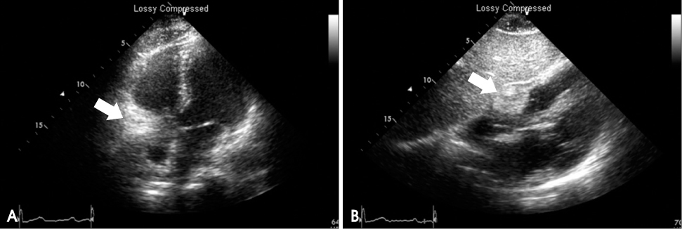
Fig. 2 Transthoracic echocardiography on the day of percutaneous coronary intervention. A: Homogeneous highly echogenic mass (white arrow) at the right atrioventricular groove was noted on apical four chamber view. B: The mass at the atrioventricular groove (white arrow) was about 3.4 cm×2.6 cm on subcostal view.
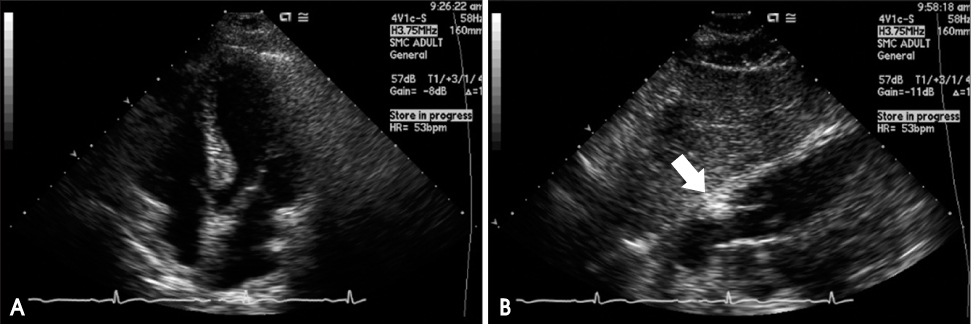
Fig. 3 Transthoracic echocardiography performed on 12 days after percutaneous coronary intervention. A: There was no visible mass at the right atrioventricular groove on apical four chamber view. B: The size of the mass (white arrow) was decreased from 3.4 cm×2.6 cm to 1.7 cm×0.7 cm on subcostal view.
Reference
-
1. Fejka M, Dixon SR, Safian RD, O'Neill WW, Grines CL, Finta B, Marcovitz PA, Kahn JK. Diagnosis, management, and clinical outcome of cardiac tamponade complicating percutaneous coronary intervention. Am J Cardiol. 2002. 90:1183–1186.
Article2. Von Sohsten R, Kopistansy C, Cohen M, Kussmaul WG 3rd. Cardiac tamponade in the "new device era": evaluation of 6999 consecutive percutaneous coronary interventions. Am Heart J. 2000. 140:279–283.
Article3. Kimbiris D, Iskandrian AS, Goel I, Bemis CE, Gehl L, Owens J, Segal BL. Transluminal coronary angioplasty complicated by coronary artery perforation. Cathet Cardiovasc Diagn. 1982. 8:481–487.
Article4. Grollier G, Bories H, Commeau P, Foucault JP, Potier JC. Coronary artery perforation during coronary angioplasty. Clin Cardiol. 1986. 9:27–29.
Article5. Topaz O, Cowley MJ, Vetrovec GW. Coronary perforation during angioplasty: angiographic detection and demonstration of complete healing. Cathet Cardiovasc Diagn. 1992. 27:284–288.
Article6. Teirstein PS, Hartzler GO. Nonoperative management of aortocoronary saphenous vein graft rupture during percutaneous transluminal coronary angioplasty. Am J Cardiol. 1987. 60:377–378.
Article7. Nassar H, Hasin Y, Gotsman MS. Cardiac tamponade following coronary arterial rupture during coronary angioplasty. Cathet Cardiovasc Diagn. 1991. 23:177–179.
Article8. Ellis SG, Ajluni S, Arnold AZ, Popma JJ, Bittl JA, Eigler NL, Cowley MJ, Raymond RE, Safian RD, Whitlow PL. Increased coronary perforation in the new device era. Incidence, classification, management, and outcome. Circulation. 1994. 90:2725–2730.
Article9. Javaid A, Buch AN, Satler LF, Kent KM, Suddath WO, Lindsay J Jr, Pichard AD, Waksman R. Management and outcomes of coronary artery perforation during percutaneous coronary intervention. Am J Cardiol. 2006. 98:911–914.
Article10. Bittl JA, Ryan TJ Jr, Keaney JF Jr, Tcheng JE, Ellis SG, Isner JM, Sanborn TA. Coronary artery perforation during excimer laser coronary angioplasty. The percutaneous excimer laser coronary angioplasty registry. J Am Coll Cardiol. 1993. 21:1158–1165.11. Klein LW. Coronary artery perforation during interventional procedures. Catheter Cardiovasc Interv. 2006. 68:713–717.
Article12. Del Campo C, Zelman R. Successful non-operative management of right coronary artery perforation during percutaneous coronary intervention in a patient receiving abciximab and aspirin. J Invasive Cardiol. 2000. 12:41–43.13. Shirakabe A, Takano H, Nakamura S, Kikuchi A, Sasaki A, Yamamoto E, Kawashima S, Takagi G, Fujita N, Aoki S, Asai K, Yoshikawa M, Kato K, Yamamoto T, Takayama M, Takano T. Coronary perforation during percutaneous coronary intervention. Int Heart J. 2007. 48:1–9.
- Full Text Links
-
- Actions
-
Cited
- CITED
-
- Close
- Share
-
- Similar articles
-
- Recent Advances in Percutaneous Coronary Intervention in Coronary Artery Disease
- Right Ventricular Wall Hematoma Secondary to Percutaneous Coronary Intervention
- A Case of Percutaneous Transcatheter Coil Embolization for Congenital Coronary Arteriovenous Fistula
- Abdominal Wall Hematoma as a Rare Complication following Percutaneous Coronary Intervention
- Forearm Compartment Syndrome after Transradial Percutaneous Coronary Artery Intervention
