Tympanometry and CT Measurement of Middle Ear Volumes in Patients with Unilateral Chronic Otitis Media
- Affiliations
-
- 1Department of Otorhinolaryngology-Head and Neck Surgery, Konkuk University School of Medicine, Seoul, Korea. hpark@kuh.ac.kr
- 2Department of Radiologys, Konkuk University School of Medicine, Seoul, Korea.
- KMID: 1486083
- DOI: http://doi.org/10.3342/ceo.2008.1.3.139
Abstract
OBJECTIVES
The goals of the study were to compare the middle ear (ME) volumes from both normal and lesioned ears, and these ME volumes were measured by a digital image processing computed tomography (CT) program in patients with unilateral chronic otitis media, and we wanted to compare the ME volumes of the lesioned ears by comparing the ME volumes obtained by tympanometry with those ME volumes measured by the digital image processing CT program.
METHODS
Forty-four patients who had unilateral chronic otitis media (COM) and contralateral normal tympanic membranes (TM) and 100 subjects with normal TMs were included in the study. The normal volumes of the external auditory canal (EAC) were measured in the normal group. The tympanometric ME volumes in the ears with a perforated TM were calculated as the difference of the tympanometric volumes measured from the both ears in patients with unilateral COM. The CT ME volumes were measured by a digital image processing program.
RESULTS
The tympanometric volumes of the EACs in the ears with normal TMs were 1.4+/-0.3 mL. There were no significant differences according to gender, age and the side of the face the ear was on. The tympanometric volumes of the EAC in the normal-side ear of the patients with unilateral COM showed no significant differences when compared with those from the normal group. The ME volumes of the intact ears, as measured by CT, showed significantly higher values than those ME volumes of the lesioned ears. The ME volumes of the lesioned ears, as measured by tympanometry, showed a strong, significant linear correlation with those ME volumes calculated by CT; however, the ME volumes of the lesioned ears, as measured by tympanometry (1.5+/-1.4 mL), were significantly larger than those ME volumes measured by CT (1.1+/-0.8 mL).
CONCLUSION
Our results show that chronic otitis media causes reduced ME volumes compared to those ME volumes of the contralateral normal ears. Tympanometry can provide a valuable estimation of the ME volumes in chronic ears, although it tends to overestimate the ME volumes, and especially for the ears with a larger ME volume.
MeSH Terms
Figure
-
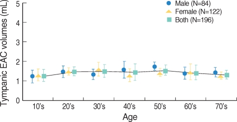
Fig. 1 The distribution of external auditory canal (EAC) volumes according to age group. There was no statistically significant difference in EAC volumes between age groups. The mean values are illustrated with error bars indicating±1 SD.

Fig. 2 Middle-ear volimes by tympanometry and CT.

Fig. 3 The correlation of the middle-ear (ME) volumes measured by CT and tympanometry. The solid lines represent a best-fit linear regression line through the data points. Linear regression analysis gives a strong correlation coefficient (R-sq=0.85), and highly significant correlation (P<0.001). The dotted lines indicate a slope of 1 (CT volumes=tympanometric volumes).
Cited by 1 articles
-
Normal Mastoid Air Cell System Geometry: Has Surface Area Been Overestimated?
Sung Wan Byun, Seung-Sin Lee, Jin Young Park, Jeong Hyun Yoo
Clin Exp Otorhinolaryngol. 2016;9(1):27-32. doi: 10.21053/ceo.2016.9.1.27.
Reference
-
1. Mehta RP, Rosowski JJ, Voss SE, O'Neil E, Merchant SN. Determinants of hearing loss in perforations of the tympanic membrane. Otol Neurotol. 2006; 2. 27(2):136–143. PMID: 16436981.
Article2. Molvaer OI, Vallersnes FM, Kringlebotn M. The size of the middle ear and the mastoid air cell. Acta Otolaryngol. 1978; Jan–Feb. 85(1-2):24–32. PMID: 626053.3. Aktas D, Kutlu R. The relationship between traumatic tympanic membrane perforations and pneumatization of the mastoid. ORL J Otorhinolaryngol Relat Spec. 2000; Nov–Dec. 62(6):311–315. PMID: 11054014.4. Vrabec JT, Champion SW, Gomez JD, Johnson RF Jr, Chaljub G. 3D CT imaging method for measuring temporal bone aeration. Acta Otolaryngol. 2002; 12. 122(8):831–835. PMID: 12542201.
Article5. Koç A, Ekinci G, Bilgili AM, Akpinar IN, Yakut H, Han T. Evaluation of the mastoid air cell system by high resolution computed tomography: three-dimensional multiplanar volume rendering technique. J Laryngol Otol. 2003; 8. 117(8):595–598. PMID: 12956911.
Article6. Lee DH, Jun BC, Kim DG, Jung MK, Yeo SW. Volume variation of mastoid pneumatization in different age groups: a study by three-dimensional reconstruction based on computed tomography images. Surg Radiol Anat. 2005; 3. 27(1):37–42. PMID: 15349696.
Article7. Shanks JE, Lilly DJ. An evaluation of tympanometric estimates of ear canal volume. J Speech Hear Res. 1981; 12. 24(4):557–566. PMID: 7329051.
Article8. Lindeman P, Holmquist J. Measurement of middle ear volume using the impedance audiometer. Am J Otol. 1981; 4. 2(4):301–303. PMID: 7258323.9. Sirikçi A, Bayazit YA, Kervancioğlu S, Ozer E, Kanlikama M, Bayram M. Assessment of mastoid air cell size versus sigmoid sinus variables with a tomography-assisted digital image processing program and morphometry. Surg Radiol Anat. 2004; 4. 26(2):145–148. PMID: 14673624.10. Voss SE, Rosowski JJ, Merchant SN, Peake WT. How do tympanic-membrane perforations affect human middle-ear sound transmission? Acta Otolaryngol. 2001; 1. 121(2):169–173. PMID: 11349771.
