J Korean Ophthalmol Soc.
2008 Jul;49(7):1041-1045. 10.3341/jkos.2008.49.7.1041.
Clinical Analysis of Fitting a Scleral Shell over Phthisis Bulbi or Discolored Blind Eyes
- Affiliations
-
- 1Department of Ophthalmology, College of Medicine, Inje University, Pusan, Korea. jwsyhyo@yahoo.co.kr
- KMID: 1476956
- DOI: http://doi.org/10.3341/jkos.2008.49.7.1041
Abstract
-
PURPOSE: To show that scleral shells can be good cosmetic prostheses for phthisis bulbi or discolored blind eyes for which no evisceration or enucleation is indicated.
METHODS
Twenty patients with phthisis bulbi or discolored blind eyes were enrolled in this study. All patients were using scleral shells. We evaluated any complications induced by the scleral shells and the cosmetic results during the follow-up period.
RESULTS
The average central thickness of the scleral shell was 1.84+/-0.26 mm, and the average volume was 1.52+/-0.25 ml. The average difference in palpebral fissure width between the fellow eye and the eye with a fitted scleral shell was 0.8+/-0.62 mm. One patient complained about ocular irritation and difficulty in fitting but was satisfied with the cosmetic appearance.
CONCLUSIONS
Fitting scleral cover shells over phthisis bulbi or discolored blind eyes without evisceration or enucleation could be a successful remedy for enhancing cosmetic appearance.
Keyword
Figure
-

Figure 1. Measuring the central thickness of the scleral shell using micrometer (Mitutoyo, Japan).
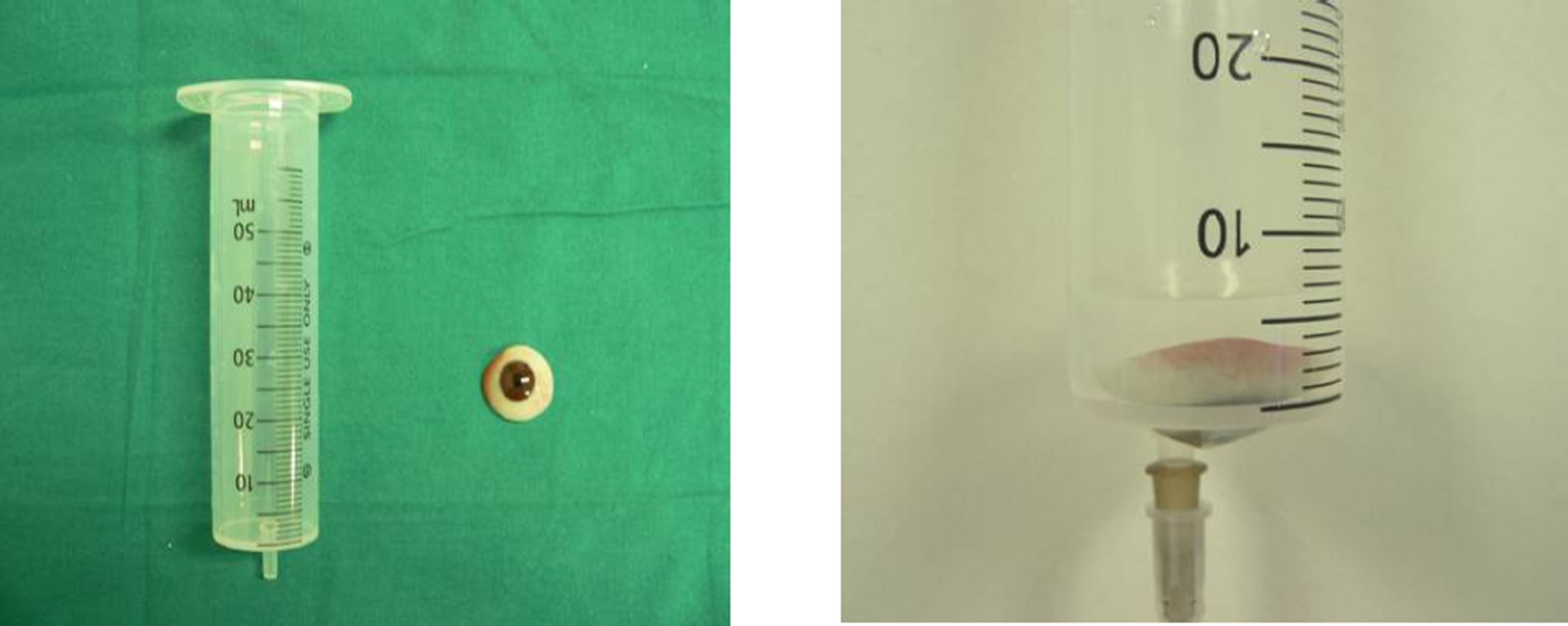
Figure 2. Measuring the volume of the scleral shell using a 50 ml single-use syringe.

Figure 3. The Scleral Shell Prosthesis: a thin hard acrylic shell-like artificial eye.

Figure 4. Photographs of a patient with phthisis bulbi. (A) Before wearing a scleral shell, he complained about ptosis and exotropia. (B) After wearing a scleral shell, he was satisfied with cosmetic appearance.
Reference
-
References
1. Bier N. Prosthetic correction. Am J Optom Physiol Opt. 1982; 59:178–83.
Article2. Spraul CW, Lang GK. Contact lenses and corneal shields. Curr Opin Ophthalmol. 1997; 8:67–75.
Article3. Buettner H, Bartley GB. Tissue breakdown and exposure associated with orbital hydroxyapatite implants. Am J Ophthalmol. 1992; 113:669–73.
Article4. Goldberg RA, Holds JB, Ebrahimpour J. Exposed hydroxyapatite orbital implants. Ophthalmology. 1992; 95:831–6.
Article5. Nunery WR, Hung GW, Bonnin JM. . Exposure rate of hydroxyapatite spheres in the anophthalmic socket: histopathologic correlation and comparison with silicone sphere implants. Ophthal Plast Reconstr Surg. 1993; 9:96–104.6. Ma'luf RN, Awwad ST. Mucous membrane graft versus Gunderson conjunctival flap for fitting a scleral shell over a sensitive cornea. Ophthal Plast Reconstr Surg. 2005; 21:356–8.7. Dortzbach RK, Woog JJ. Choice of procedure Enucleation, evisceration, or prosthetic fitting over globes. Ophthalmology. 1985; 92:1249–55.8. Chung WS, Kim CJ. Clinical Experience of Hydroxyapatite Orbital Implant. J Korean Ophthalmol Soc. 1994; 35:615–21.9. Fahim DK, Frueh BR, Musch DC, Nelson CC. Complications of pegged and non-pegged hydroxyapatite orbital implants. Ophthal Plast Reconstr Surg. 2007; 23:206–10.
Article10. Lin CJ, Liao SL, Jou JR. . Complications of motility peg placement for porous hydroxyapatite orbital implants. Br J Ophthalmol. 2002; 86:394–6.
Article11. Bailey CS, Buckley RJ. Ocular prostheses and contact lenses I-Cosmetic devices. BMJ. 1991; 302:1010–2.
Article12. Cote RE, Haddad SE. Fitting a prosthesis over phthisis bulbi or discolored blind eyes. Adv Ophthalmic Plast Reconstr Surg. 1990; 8:136–45.13. Thaller VT. Enucleation volume measurement. Ophthal Plast Reconstr Surg. 1997; 13:18–20.
Article14. Lee EY, Choi JH, Baek SH. Clinical Analysis of the Size of the Orbital Implant and Prosthesis in Eviscerated Patients. J Korean Ophthalmol Soc. 2003; 44:1254–9.15. Kaltreider SA. The ideal ocular prosthesis. Ophthal Plast Reconstr Surg. 2000; 16:388–92.
Article
- Full Text Links
-
- Actions
-
Cited
- CITED
-
- Close
- Share
-
- Similar articles
-
- A Case of Ossification in the Phthisis Bulbi
- Preoperative Axial Length Measured by Ultrasonography in Phthisis Bulbi
- Evisceration Using Scleral Capping Technique for Severe Phthisis Bulbi
- Comparison of Evisceration with Primary Orbital Implant Surgery in Endophthalmitis and Phthisis Bulbi
- Guide for Successful Scleral Lens Fitting
