Clinical Significance of Left Ventricular Torsional Parameters during Supine Bicycle Cardiopulmonary Exercise Echocardiography
- Affiliations
-
- 1Division of Cardiology, Department of Internal Medicine, Gachon University Gil Hospital, Incheon, Korea. heart@gilhospital.com
- KMID: 1473759
- DOI: http://doi.org/10.4250/jcu.2009.17.1.2
Abstract
- BACKGROUND
Left ventricular (LV) torsion plays an important role in both LV systolic and diastolic function. Notwithstanding the fact that speckle tracking imaging echocardiography (STI) is a validated method to measure LV torsion, few data regarding the clinical significance of LV torsional parameters using STI on exercise capacity during exercise echocardiography were reported.
METHODS
Fifty four participants completed the supine bicycle cardiopulmonary exercise echocardiography under a symptom-limited protocol. LV torsion was defined as the net difference between LV peak apical rotation, and basal rotation divided by LV diastolic longitudinal length. LV basal, and apical short-axis rotations at each stage were analyzed by STI.
RESULTS
LV torsion measurement was feasible in 43/54 (80%) at peak exercise. The LV torsions were increased during exercise, and even until the recovery. Peak twisting, and untwisting velocities were significantly increased during exercise, but were decreased at recovery. As expected, baseline torsion was positively correlated with LV ejection fraction and baseline apical peak untwisting velocity has correlation with E/E' (r=0.50, p<0.01 and r=0.30, p<0.05, respectively). Interestingly, apical peak twisting velocity at peak exercise was significantly correlated with maximal O2 consumption and VO2 interval change (r=0.50, p<0.01 and r=0.33, p<0.05, respectively).
CONCLUSION
It was feasible to measure LV torsion by STI at every step during exercise echocardiography, although the feasibility was relatively low at peak exercise. LV torsional parameters during exercise showed significant relations with exercise capacity as well as LV systolic and diastolic functions.
Figure
-
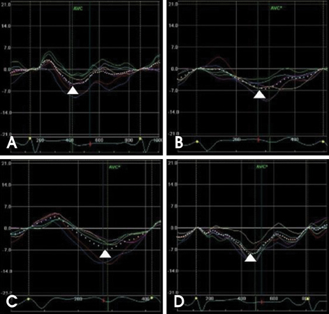
Fig. 1 Basal rotation during exercise and recovery phase. The white arrowheads represent each peak values. At baseline (A), 75 W exercise (B), peak exercise (C) and recovery (D), each basal rotation represents negative value (clockwise rotation) and increases during exercise and even till the recovery phase.
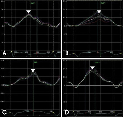
Fig. 2 Apical rotation during exercise and recovery phase. The white arrowheads represent each peak values. At baseline (A), 75 W exercise (B), peak exercise (C) and recovery (D), each apical rotation represents positive value (counterclockwise rotation) and increases during exercise and even till the recovery phase.
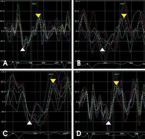
Fig. 3 Basal peak twisting and untwisting velocities during exercise and recovery phase. The white arrowheads represent each peak twisting velocities and yellow arrowheads represent each peak untwisting velocities. At baseline (A), 75 W exercise (B), peak exercise (C) and recovery (D), both twisting and untwisting velocity were increased during exercise and decreased at recovery phase.

Fig. 4 Apical peak twisting and untwisting velocity during exercise and recovery phase. The white arrowheads represent each peak twisting velocities and yellow arrowheads represent each peak untwisting velocities. At baseline (A), 75 W exercise (B), peak exercise (C) and recovery (D), both twisting and untwisting velocity were increased during exercise and decreased at recovery phase.

Fig. 5 The correlation between torsional parameters at baseline and left ventricular functional parameters. There were positive correlations between torsion at baseline and left ventricular ejection fraction (r=0.50, p=0.001) (A). Apical peak untwisting velocity at baseline showed positive correlation with E/E' (r=0.30, p<0.05) (B).

Fig. 6 LV torsion (A), apical peak twisting velocity (B) and apical peak untwisting velocity (C) during exercise echocardiography. LV torsion increases during exercise and recovery. However, apical peak twisting velocity increases during exercise and decreases at recovery phase and also absolute value of apical peak untwisting velocity increases during exercise and decreases at recovery phase.
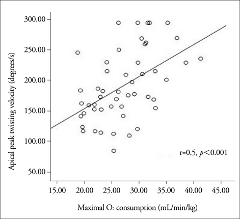
Fig. 7 The correlation between maximal O2 consumption and apical peak twisting velocity at peak exercise. Maximal O2 consumption (MVO2, mL/min/kg) correlated with apical peak twisting velocity at peak exercise (r=0.5, p<0.001).
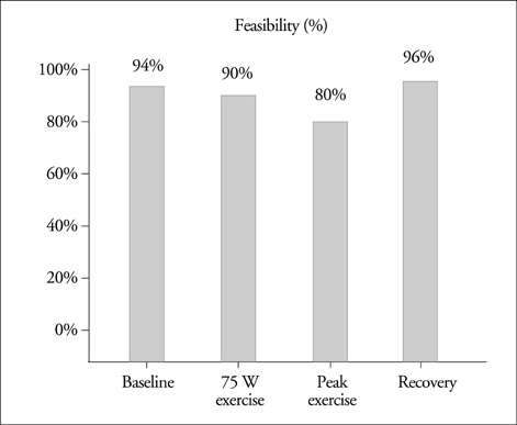
Fig. 8 The Feasibility of left ventricular torsion. Measurements of left ventricular torsion were feasible in 51/54 (94%) at baseline, 49/54 (90%) at 75 W exercise, 43/54 (80%) at peak exercise and 52/54 (96%) at recovery, respectively.
Reference
-
1. Rothfeld JM, LeWinter MM, Tischler MD. Left ventricular systolic torsion and early diastolic filling by echocardiography in normal humans. Am J Cardiol. 1998. 81:1465–1469.
Article2. Notomi Y, Maureen G, Martin-Miklovic MG, Oryszak SJ, Shiota T, Deserranno D, Popovic ZB, Garcia MJ, Greenberg NL, Thomas JD. Enhanced ventricular untwisting during exercise a mechanistic manifestation of elastic recoil described by Doppler tissue imaging. Circulation. 2006. 113:2524–2533.3. Takeuchi M, Nishikage T, Nakai H, Kokumai M, Otani S, Lang RM. The assessment of left ventricular twistin anterior wall myocardial infarction using two-dimensional speckle tracking imaging. J Am Soc Echocardiogr. 2007. 20:36–44.
Article4. Tischler M, Niggel J. Left ventricular systolic torsion and exercise in normal hearts. J Am Soc Echocardiogr. 2003. 16:670–674.
Article5. Notomi Y, Setser RM, Shiota T, Martin-Miklovic MG, Weaver JA, Popović ZB, Yamada H, Greenberg NL, White RD, Thomas JD. Assessment of left ventricular torsional deformation by Doppler tissue imaging validation study with tagged magnetic resonance imaging. Circulation. 2005. 111:1141–1147.
Article6. Notomi Y, Lysyansky P, Setser RM, Shiota T, Popovic ZB, Martin-Miklovic MG, Weaver JA, Oryszak SJ, Greenberg NL, White RD, Thomas JD. Measurement of ventricular torsion by two-dimensional ultrasound speckle tracking imaging. J Am Coll Cardiol. 2005. 45:2034–2041.
Article7. Helle-Valle T, Crosby J, Edvardsen T, Lyseggen E, Amundsen BH, Smith HJ, Rosen BD, Lima JAC, Torp H, Ihlen H, Smiseth OA. New noninvasive method for assessment of left ventricular rotation speckle tracking echocardiography. Circulation. 2005. 112:3149–3156.
Article8. Nakai H, Takeuchi M, Nishikage T, Kokumai M, Otani S, Lang RM. Effect of aging on twist-displacement loop by 2-dimensional speckle tracking imaging. J Am Soc Echocardiogr. 2006. 19:880–885.
Article9. Takeuchi M, Nakai H, Kokumai M, Nishikage T, Otani S, Lang RM. Age-related changes in left ventricular twist assessed by two-dimensional speckle-tracking imaging. J Am Soc Echocardiogr. 2006. 19:1077–1084.
Article10. Kim HK, Sohn DW, Lee SE, Choi SY, Park JS, Kim YJ, Oh BH, Park YB, Choi YS. Assessment of left ventricular rotation and torsion with twodimensional speckle tracking echocardiography. J Am Soc Echocardiogr. 2007. 20:45–53.
Article11. Reuss CS, Moreno CA, Appleton CP, Lester SJ. Doppler tissue imaging during supine and upright exercise in healthy adults. J Am Soc Echocardiogr. 2005. 18:1343–1348.
Article12. Thomas JD, Picard MH, Yoerger DM, Wood MJ. Myocardial adaptation to short-term high-intensity exercise in highly trained athletes. J Am Soc Echocardiogr. 2006. 19:1280–1285.
Article13. Yun KL, Niczyporuk MA, Daughters GT, Ingels NB, Stinson EB, Alderman EL, Hansen DE, Miller DC. Alterations in left ventricular diastolic twist mechanics during acute human cardiac allograft rejection. Circulation. 1991. 83:962–973.
Article14. Davis KL, Mehlhorn U, Schertel ER, Geissler HJ, Trevas D, Laine GA, Allen SJ. Variation in tau, the time constant for isovolumic relaxation, along the left ventricular base-to-apex axis. Basic Res Cardiol. 1999. 94:41–48.
Article15. Ommen SR, Nishimura RA, Appleton CP, Miller FA, Oh JK, Redfield MM, Tajik AJ. Clinical utility of Doppler echocardiography and tissue Doppler imaging in the estimation of left ventricular filling pressures: a comparative simultaneous Doppler-catheterization study. Circulation. 2000. 102:1788–1794.
Article16. D'Souza KA, Mooney DJ, Russell AE, MacIsaac AI, Aylward PE, Prior DL. Abnormal septal motion affects early diastolic velocities at the septal and lateral mitral annulus, and impacts on estimation of the pulmonary capillary wedge pressure. J Am Soc Echocardiogr. 2005. 18:445–453.
- Full Text Links
-
- Actions
-
Cited
- CITED
-
- Close
- Share
-
- Similar articles
-
- Evaluation of Left Ventricular Function by Dynamic Exercise Echocardiography in Normal Subjects
- The Usefulness of Supine Bycycle Stress Echocardiography for Evaluationg Coronary Artery Disease: Comparision with Exercise Electrocardiography
- The Time Course and Determinants of B-Type Natriuretic Peptide in Healthy Men during Supine Bicycle Exercise
- The Association of Left Ventricular Hypertrophy with Intraventricular Dyssynchrony at Rest and during Exercise in Hypertensive Patients
- Exercise Echocardiography in Asymptomatic Patients with Severe Aortic Stenosis and Preserved Left Ventricular Ejection Fraction
