J Cardiovasc Ultrasound.
2009 Dec;17(4):151-152. 10.4250/jcu.2009.17.4.151.
An Unusual Presentation of an Atrial Septal Defect
- Affiliations
-
- 1The Heart Center of Chonnam National University Hospital, Gwangju, Korea. jcpark54@hanmail.net
- KMID: 1473721
- DOI: http://doi.org/10.4250/jcu.2009.17.4.151
Abstract
- No abstract available.
MeSH Terms
Figure
-

Fig. 1 Transthoracic echocardiography showed 0.97 cm sized atrial septal defect suspected to be sinus venosus type. Left atrium looked like divided chambers by abnormal membranous structure.
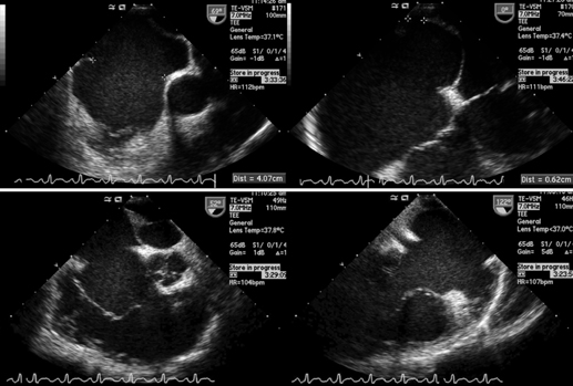
Fig. 2 Transesophageal echocardiography demonstrated 4.07 cm sized large atrial septal defect and membranous structures divided left atrium into plural chambers. Therefore, it made atrial chambers of large right atrium and small left atrium.
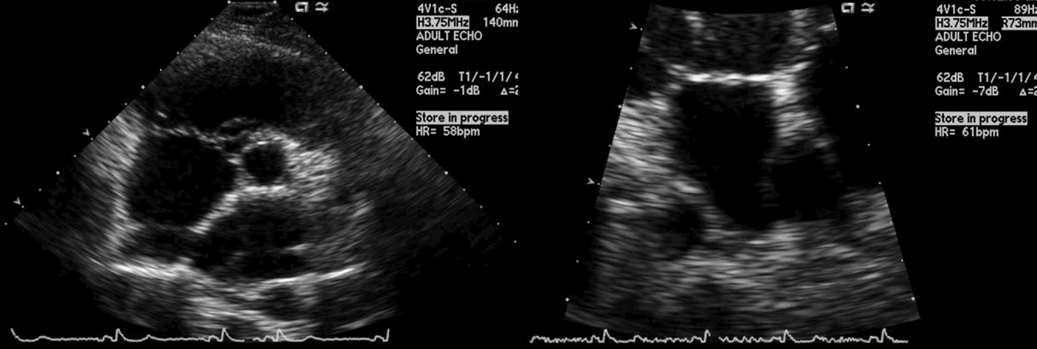
Fig. 3 Follow-up transthoracic echocardiography showed no remnant atrial septal defect, but left atrium still looked like divided chambers by membranous structure.
Reference
-
1. Sharon IC. Atrial septal deftect in the adult. J Natl Med Assoc. 1987. 79:1095–1097.2. Gutgesell HP, Huhta JC, Latson LA, Huffines D, McNamara DG. Accuracy of two-dimensional echocardiography in the diagnosis of congenital heart disease. Am J Cardiol. 1985. 55:514–518.
Article3. Feigenbaum H, Armstrong WF, Ryan T. Feigenbaum's echocardiography: Congenital heart diseases. 2005. 6th ed. Philadelphia: Lippincott Williams and Wilkins;559–636.
- Full Text Links
-
- Actions
-
Cited
- CITED
-
- Close
- Share
-
- Similar articles
-
- A Case of Atrial Septal Defect in Identical Twins
- A Case of Atrial Septal Aneurysm Associated with Atrial Septal Defect
- Simultaneous Closure of a Left Atrial Appendage through an Atrial Septal Defect and the Atrial Septal Defect
- Transcatheter Closure of Secundum Atrial Septal Defect with the Amplatzer Septal Occluder
- Procedural, Early and Long-Term Outcomes after Transcatheter Atrial Septal Defects Closure: Comparison between Large and Very Large Atrial Septal Defect Groups
