Comparison of model analysis measurements among plaster model, laser scan digital model, and cone beam CT image
- Affiliations
-
- 1Department of Orthodontics, School of Dentistry, Chosun University, Korea. shlim@chosun.ac.kr
- KMID: 1469134
- DOI: http://doi.org/10.4041/kjod.2009.39.1.6
Abstract
OBJECTIVE
The purpose of this study was to evaluate the possibility of using a digital model and cone beam computed tomograph (CBCT) image for model analysis.
METHODS
Model analyses of CBCT images, plaster models, and digital models of 20 orthodontic patients with a permanent dentition with no proximal metal restorations, were compared.
RESULTS
The average differences of tooth size measurements were 0.01 to 0.20 mm, and the average difference of arch length discrepancy measurements were 0.41 mm in the maxilla and 0.82 mm in the mandible. The difference in Bolton discrepancy measurements was 0.17 mm for the anterior region and 0.44 mm overall but with no statistically significant difference. When comparing CBCT images with plaster models, the average differences in tooth size measurements were -0.22 to 0.01 mm, and the average differences in arch length discrepancy measurements were 0.43 mm in the maxilla and 0.32 mm in the mandible. Difference in Bolton discrepancy measurements were 0.35 mm in the anterior region and 1.25 mm overall. CBCT images showed significantly smaller overall Bolton discrepancy measurements.
CONCLUSIONS
Although there were statistically significant differences in some model analysis measurements, the ranges of measurement errors of the digital model and CBCT images were clinically acceptable. Therefore, a digital model and CBCT image can be used for model analysis.
Keyword
Figure
-
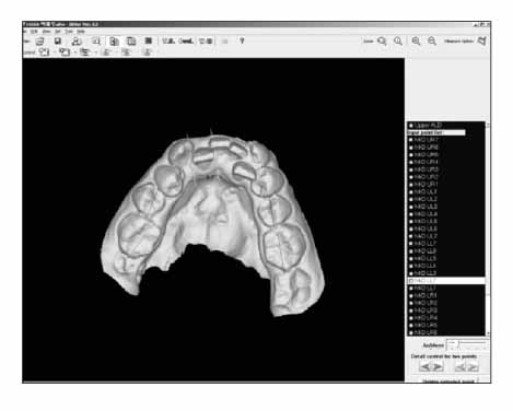
Fig 1 Measurements of tooth size and dental arch width of a laser scan digital model. 3DXer ver 3.5 was used.
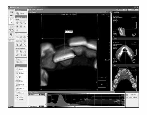
Fig 2 Volume rendering model of CBCT data constructed using Accurex ver 1.0. 3D zoom mode was applied and displayed in the main window for measurement of lower left lateral incisor width. The thresholding ranges of teeth 3 preset that were used in this study are shown in the fine tuning tab on the bottom of the program window.
Cited by 8 articles
-
Comparison of bony changes between panoramic radiograph and cone beam computed tomographic images in patients with temporomandibular joint disorders
Dong-Yul Lee, Yun-Jung Kim, Yun-Heon Song, Nam-Ho Lee, Yong-Kyu Lim, Sung-Taek Kang, Sug-Joon Ahn
Korean J Orthod. 2010;40(6):364-372. doi: 10.4041/kjod.2010.40.6.364.Validity of palatal superimposition of 3-dimensional digital models in cases treated with rapid maxillary expansion and maxillary protraction headgear
Jin-Il Choi, Bong-Kuen Cha, Paul-Georg Jost-Brinkmann, Dong-Soon Choi, In-San Jang
Korean J Orthod. 2012;42(5):235-241. doi: 10.4041/kjod.2012.42.5.235.Comparison of arch form between ethnic Malays and Malaysian Aborigines in Peninsular Malaysia
Siti Adibah Othman, Eunice Soh Xinwei, Sheh Yinn Lim, Marhazlinda Jamaludin, Nor Himazian Mohamed, Zamros Yuzaidi Mohd Yusof, Lily Azura Shoaib, Nik Noriah Nik Hussein
Korean J Orthod. 2012;42(1):47-54. doi: 10.4041/kjod.2012.42.1.47.New classification of lingual arch form in normal occlusion using three dimensional virtual models
Kyung Hee Park, Mohamed Bayome, Jae Hyun Park, Jeong Woo Lee, Seung-Hak Baek, Yoon-Ah Kook
Korean J Orthod. 2015;45(2):74-81. doi: 10.4041/kjod.2015.45.2.74.Clinical application of an intraoral scanner for serial evaluation of orthodontic tooth movement: A preliminary study
Dalsun Yun, Dong-Soon Choi, Insan Jang, Bong-Kuen Cha
Korean J Orthod. 2018;48(4):262-267. doi: 10.4041/kjod.2018.48.4.262.Three-dimensional comparison of 2 digital models obtained from cone-beam computed tomographic scans of polyvinyl siloxane impressions and plaster models
Jin-Yi Park, Dasomi Kim, Sang-Sun Han, Hyung-Seog Yu, Jung-Yul Cha
Imaging Sci Dent. 2019;49(4):257-263. doi: 10.5624/isd.2019.49.4.257.Maxillomandibular arch width differences at estimated centers of resistance: Comparison between normal occlusion and skeletal Class III malocclusion
Yun-Jin Koo, Sung-Hwan Choi, Byeong-Tak Keum, Hyung-Seog Yu, Chung-Ju Hwang, Birte Melsen, Kee-Joon Lee
Korean J Orthod. 2017;47(3):167-175. doi: 10.4041/kjod.2017.47.3.167.Comparison of midsagittal reference plane in PA cephalogram and 3D CT
Jin-Hyoung Cho, Ji-Yeon Moon
Korean J Orthod. 2010;40(1):6-15. doi: 10.4041/kjod.2010.40.1.6.
Reference
-
1. OrthoCAD. USA: Available at: http://www.orthocad.com.2. Emodels. USA: Available at: http://www.dentalemodels.com.3. Zilberman O, Huggare JA, Parikakis KA. Evaluation of the validity of tooth size and arch width measurements using conventional and three-dimensional virtual orthodontic models. Angle Orthod. 2003. 73:301–306.4. Kuroda T, Motohashi N, Tominaga R, Iwata K. Three-dimensional dental cast analyzing system using laser scanning. Am J Orthod Dentofacial Orthop. 1996. 110:365–369.
Article5. Sohmura T, Kojima T, Wakabayashi K, Takahashi J. Use of an ultrahigh-speed laser scanner for constructing three-dimensional shapes of dentition and occlusion. J Prosthet Dent. 2000. 84:345–352.
Article6. Stevens DR, Flores-Mir C, Nebbe B, Raboud DW, Heo G, Major PW. Validity, reliability, and reproducibility of plaster vs digital study models: comparison of peer assessment rating and Bolton analysis and their constituent measurements. Am J Orthod Dentofacial Orthop. 2006. 129:794–803.
Article7. Garino F, Garino GB. Comparison of dental arch measurements between stone and digital casts. World J Orthod. 2002. 3:250–254.8. Baik HS, Lee HJ, Jeon JM. A proposal of soft tissue landmarks for craniofacial analysis using three-dimensional laser scan imaging. Korean J Orthod. 2006. 36:1–13.9. Baik HS, Jeon JM, Lee HJ. A study of facial soft tissue of Korean adults with normal occlusion using a three-dimensional laser scanner. Korean J Orthod. 2006. 36:14–29.10. Ko SD, Cha KS. A Study on the labial & buccal surface contour in Korean permanent teeth using three-dimensional laser scanning. Korean J Orthod. 2002. 32:275–291.11. Chang YJ, Kim TW, Yoo KH. The effect of variations in the vertical position of the bracket on the crown inclination. Korean J Orthod. 2002. 32:401–411.12. You HW. The optimal scanning pose generating algorithm for 3D dental model scanner [thesis]. 2000. Seoul: Myongji University.13. Park JW. The study on the measurement error of the 3D-digital model made by laser scan method [thesis]. 2003. Seoul: Seoul National University.14. Han JH. Comparison of measurements in three-dimensional digital model and dental plaster model [thesis]. 2004. Seoul: Yonsei University.15. Park SI. The comparison between manual and 3D-digital measurement in dental cast measurements according to the degree of crowding [thesis]. 2006. Seoul: Korea University.16. Holberg C, Steinhäuser S, Geis P, Rudzki-Janson I. Conebeam computed tomography in orthodontics: benefits and limitations. J Orofac Orthop. 2005. 66:434–444.
Article17. Mischkowski RA, Pulsfort R, Ritter L, Neugebauer J, Brochhagen HG, Keeve E, et al. Geometric accuracy of a newly developed cone-beam device for maxillofacial imaging. Oral Surg Oral Med Oral Pathol Oral Radiol Endod. 2007. 104:551–559.
Article18. Chae JH, Song JW, Cha JY, Choi JS, Park YC. Labial and buccal surface contours of Korean normal occlusion in a three-dimensional digital model. Korean J Orthod. 2008. 38:95–103.
Article19. Misch KA, Yi ES, Sarment DP. Accuracy of cone beam computed tomography for periodontal defect measurements. J Periodontol. 2006. 77:1261–1266.
Article20. Pinsky HM, Dyda S, Pinsky RW, Misch KA, Sarment DP. Accuracy of three-dimensional measurements using cone-beam CT. Dentomaxillofac Radiol. 2006. 35:410–416.
Article21. Ludlow JB, Laster WS, See M, Bailey LJ, Hershey HG. Accuracy of measurements of mandibular anatomy in cone beam computed tomography images. Oral Surg Oral Med Oral Pathol Oral Radiol Endod. 2007. 103:534–542.
Article22. Chang J, Yenice KM, Narayana A, Gutin PH. Accuracy and feasibility of cone-beam computed tomography for stereotactic radiosurgery setup. Med Phys. 2007. 34:2077–2084.
Article23. Bolton WA. Disharmony in tooth size and its relation to the analysis and treatment of malocclusion. Angle Orthod. 1958. 28:113–130.24. Lascala CA, Panella J, Marques MM. Analysis of the accuracy of linear measurements obtained by cone beam computed tomography (CBCT-NewTom). Dentomaxillofac Radiol. 2004. 33:291–294.
Article25. Santoro M, Galkin S, Teredesai M, Nicolay OF, Cangialosi TJ. Comparison of measurements made on digital and plaster models. Am J Orthod Dentofacial Orthop. 2003. 124:101–105.
Article26. Quimby ML, Vig KW, Rashid RG, Firestone AR. The accuracy and reliability of measurements made on computer-based digital models. Angle Orthod. 2004. 74:298–303.27. Lee JH, Paik KS, Chang MS, Lee SP. An evaluation of validity of measurements using digital caliper and three-dimensional virtual dental models. Korean J Anat. 2004. 37:209–218.28. Bolton WA. The clinical application of tooth-size analysis. Am J Orthod. 1962. 48:504–529.29. Tomassetti JJ, Taloumis LJ, Denny JM, Fischer JR Jr. A comparison of 3 computerized Bolton tooth-size analyses with a commonly used method. Angle Orthod. 2001. 71:351–357.30. Lagravère MO, Fang Y, Carey J, Toogood RW, Packota GV, Major PW. Density conversion factor determined using a cone-beam computed tomography unit NewTom QR-DVT 9000. Dentomaxillofac Radiol. 2006. 35:407–409.
Article31. Sim EJ, Hwang HS, Moon JD. A study on the error of tooth size measurements. Korean J Orthod. 1999. 29:491–501.32. Anatomage. USA: Available at: http://www.anatomage.com.33. OrthoProofUSA. USA: Available at: http://www.orthoproofusa.com.
- Full Text Links
-
- Actions
-
Cited
- CITED
-
- Close
- Share
-
- Similar articles
-
- Accuracy and reliability of measurements performed using two different software programs on digital models generated using laser and computed tomography plaster model scanners
- Three-dimensional comparison of 2 digital models obtained from cone-beam computed tomographic scans of polyvinyl siloxane impressions and plaster models
- The reliability of the cephalogram generated from cone-beam CT
- A method for mandibular dental arch superimposition using 3D cone beam CT and orthodontic 3D digital model
- Accuracy of Bolton analysis measured in laser scanned digital models compared with plaster models (gold standard) and cone-beam computer tomography images
