Assessment of Insulin Resistance and Its Clinical Application
- Affiliations
-
- 1Department of Internal Medicine, Graduate School of Medicine, Gachon University of Medicine and Science, Korea.
- 2Laboratory of Molecular Endocrinology, Graduate School of Medicine, Gachon University of Medicine and Science, Korea.
- KMID: 1468500
- DOI: http://doi.org/10.3803/jkes.2009.24.2.75
Abstract
- No abstract available.
MeSH Terms
Figure
-
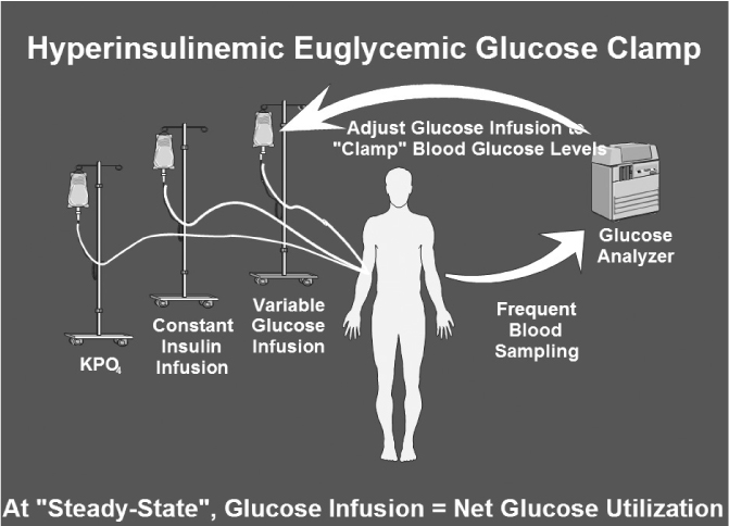
Fig. 1 Hyperinsulinemic Euglycemic Glucose Clamp Technique. After an overnight fast, insulin is infused intravenously at a constant rate that may range from 5~120 mU/m2/min (dose per body surface area per minute). This constant insulin infusion results in a new steady-state insulin level that is above the fasting level (hyperinsulinemic). As a consequence, glucose disposal in skeletal muscle and adipose tissue is increased while hepatic glucose production is suppressed. Under these conditions, a bedside glucose analyzer is used to frequently monitor blood glucose levels at 5~10 min intervals while 20% dextrose is given intravenously at a variable rate in order to "clamp" blood glucose concentrations in the normal range (euglycemic). An infusion of potassium phosphate is also given to prevent hypokalemia resulting from hyperinsulinemia and increased glucose disposal. After several hours of constant insulin infusion, steady-state conditions can typically be achieved for plasma insulin, blood glucose, and the glucose infusion rate (GIR). Assuming that the hyperinsulinemic state is sufficient to completely suppress hepatic glucose production, and since there is no net change in blood glucose concentrations under steady-state clamp conditions, the GIR must be equal to the glucose disposal rate (M). Thus, whole body glucose disposal at a given level of hyperinsulinemia can be directly determined (Adopted from Muniyappa R, et al. Am J Physiol Endocrinol Metab 294:E15-26, 2008).
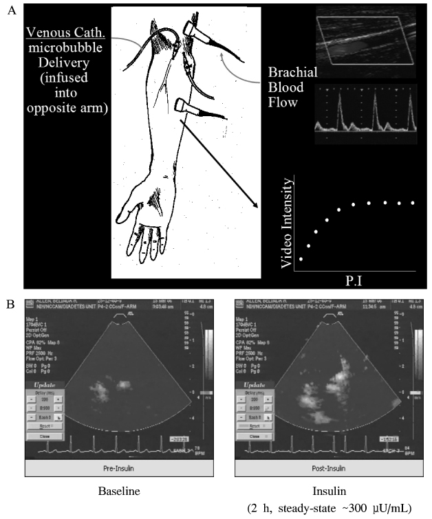
Fig. 2 Brachial artery measurements and insulin-stimulated capillary recruitment measured by contrast enhanced ultrasonography. A. Brachial artery measurements are made ~10 cm proximal to the antecubital fold while the patient is in the supine position using a high frequency L12-5 linear array transducer interfaced to an HDI5000 ultrasound (Philips Medical Systems, Andover, MA). Artery diameter is measured using 2-D imaging of the longitudinal artery as the distance between each inside edge of the arterial intima. Velocity is determined using pulse-wave Doppler and brachial flow calculated from the diameter and velocity measurements. Contrast ultrasound measurements are performed using an HDI5000 (Philips Medical Systems) while the patient is sitting upright. A P4-2 phased array transducer (Philips Medical Systems) is positioned with a ring stand and clamp to image the flexor muscles of the forearm in cross section. Microbubbles (Bristol-Myers Squibb, Princeton, NJ) are delivered intravenously at a rate that produced moderate opacification (below saturation level) at the longest pulsing interval used (16s). Microbubbles are detected by power Doppler imaging using a mechanical index capable of destroying all bubbles in the ultrasound beam. After an initial equilibration period, a pulsing interval (time) versus video-intensity curve is generated. The pulsing interval is the time between successive ultrasound pulses, which destroy microbubbles, and video intensity is the intensity of signal generated from the microbubbles. At a low pulsing interval (e.g., 500 ms) only microbubbles in the larger arteries and arterioles are filled before another destructive pulse is delivered, hence all of signal intensity is secondary to filling of these vessels. At a longer pulsing interval (e.g., 16 s) the microbubbles will again have filled larger arteries and arterioles but will have also entered the microvasculature. Subtracting images obtained at 500 ms from those taken at 16 s eliminates signals generated from the background tissue or larger vessels, leaving a measure of MBV. B. Images are recorded onto SVHS videotape, captured using Adobe Premiere software (Version 6.0) and analyzed using MCE software (University of Virginia) (Adopted from Clerk LH, et al. Diabetes 55:1436-1442, 2006).
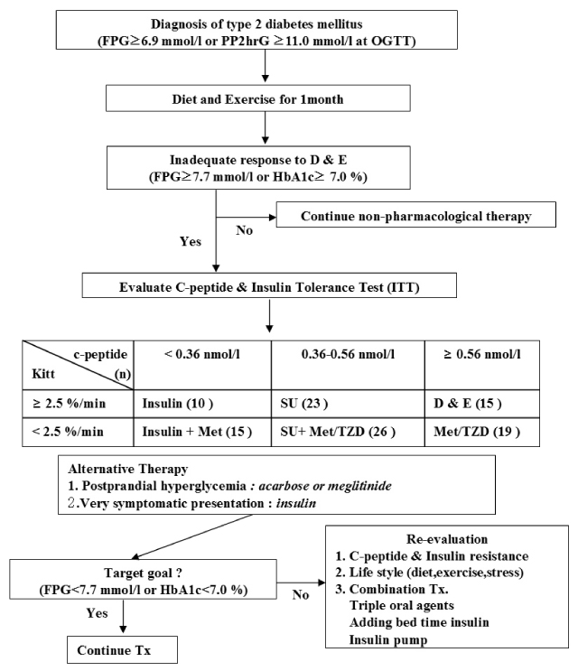
Fig. 3 New standardized therapeutic model in Korean patients with type 2 diabetes mellitus. Patients are allocated to one of six groups based on the reference value of C-peptide and the rate constant for plasma glucose disappearance (Kitt) as follows: (i) diet and exercise only; (ii) patients treated with metformin (Met) and/or thiazolidinediones (Met/TZD); (iii) patients treated with sulphonylurea only (SU); (iv) patients treated with SU + Met/TZD; (v) patients treated with insulin only; and (vi) patients treated with insulin + Met. Kitt (%/min), rate constant for glucose disappearance by the insulin tolerance test (ITT); FPG, fasting plasma glucose concentration; PP2hG, postprandial 2-h plasma glucose concentration (Adopted from Choi SH, et al.. Clin Endocrinol (Oxf.) 69:549-555, 2008).
Reference
-
1. Banting FG, Best CH. The internal secretion of the pancreas 1922. Indian J Med Res. 2007. 125:251–266.2. Muniyappa R, Montagnani M, Koh KK, Quon MJ. Cardiovascular actions of insulin. Endocr Rev. 2007. 28:463–491.3. Himsworth H. Diabetes mellitus: a differentiation into insulin-sensitive and insulin-insensitive types. Lancet. 1936. 1:127–130.4. Reaven GM. The insulin resistance syndrome: definition and dietary approaches to treatment. Annu Rev Nutr. 2005. 25:391–406.5. Geiss LS, Pan L, Cadwell B, Gregg EW, Benjamin SM, Engelgau MM. Changes in incidence of diabetes in U.S. adults, 1997-2003. Am J Prev Med. 2006. 30:371–377.6. DeFronzo RA, Tobin JD, Andres R. Glucose clamp technique: a method for quantifying insulin secretion and resistance. Am J Physiol. 1979. 237:E214–E223.7. Campbell PJ, Mandarino LJ, Gerich JE. Quantification of the relative impairment in actions of insulin on hepatic glucose production and peripheral glucose uptake in non-insulin-dependent diabetes mellitus. Metabolism. 1988. 37:15–21.8. Gelfand RA, Barrett EJ. Effect of physiologic hyperinsulinemia on skeletal muscle protein synthesis and breakdown in man. J Clin Invest. 1987. 80:1–6.9. Petersen KF, Dufour S, Befroy D, Garcia R, Shulman GI. Impaired mitochondrial activity in the insulin-resistant offspring of patients with type 2 diabetes. N Engl J Med. 2004. 350:664–671.10. Clerk LH, Vincent MA, Jahn LA, Liu Z, Lindner JR, Barrett EJ. Obesity blunts insulin-mediated microvascular recruitment in human forearm muscle. Diabetes. 2006. 55:1436–1442.11. Morris AD, Ueda S, Petrie JR, Connell JM, Elliott HL, Donnelly R. The euglycaemic hyperinsulinaemic clamp: an evaluation of current methodology. Clin Exp Pharmacol Physiol. 1997. 24:513–518.12. Shen SW, Reaven GM, Farquhar JW. Comparison of impedance to insulin-mediated glucose uptake in normal subjects and in subjects with latent diabetes. J Clin Invest. 1970. 49:2151–2160.13. Harano Y, Hidaka H, Takatsuki K, Ohgaku S, Haneda M, Motoi S, Kawagoe K, Shigeta Y, Abe H. Glucose, insulin, and somatostatin infusion for the determination of insulin sensitivity in vivo. Metabolism. 1978. 27:1449–1452.14. Bergman RN, Ider YZ, Bowden CR, Cobelli C. Quantitative estimation of insulin sensitivity. Am J Physiol. 1979. 236:E667–E677.15. Dalla Man C, Campioni M, Polonsky KS, Basu R, Rizza RA, Toffolo G, Cobelli C. Two-hour seven-sample oral glucose tolerance test and meal protocol: minimal model assessment of beta-cell responsivity and insulin sensitivity in nondiabetic individuals. Diabetes. 2005. 54:3265–3273.16. Matthews DR, Hosker JP, Rudenski AS, Naylor BA, Treacher DF, Turner RC. Homeostasis model assessment: insulin resistance and beta-cell function from fasting plasma glucose and insulin concentrations in man. Diabetologia. 1985. 28:412–419.17. Katz A, Nambi SS, Mather K, Baron AD, Follmann DA, Sullivan G, Quon MJ. Quantitative insulin sensitivity check index: a simple, accurate method for assessing insulin sensitivity in humans. J Clin Endocrinol Metab. 2000. 85:2402–2410.18. Mather KJ, Hunt AE, Steinberg HO, Paradisi G, Hook G, Katz A, Quon MJ, Baron AD. Repeatability characteristics of simple indices of insulin resistance: implications for research applications. J Clin Endocrinol Metab. 2001. 86:5457–5464.19. Chen H, Sullivan G, Yue LQ, Katz A, Quon MJ. QUICKI is a useful index of insulin sensitivity in subjects with hypertension. Am J Physiol Endocrinol Metab. 2003. 284:E804–E812.20. Chen H, Sullivan G, Quon MJ. Assessing the predictive accuracy of QUICKI as a surrogate index for insulin sensitivity using a calibration model. Diabetes. 2005. 54:1914–1925.21. Hanley AJ, Williams K, Gonzalez C, D'Agostino RB Jr, Wagenknecht LE, Stern MP, Haffner SM. San Antonio Heart Study. Mexico City Diabetes Study. Insulin Resistance Atherosclerosis study. Prediction of type 2 diabetes using simple measures of insulin resistance: combined results from the San Antonio Heart Study, the Mexico City Diabetes Study, and the Insulin Resistance Atherosclerosis Study. Diabetes. 2003. 52:463–469.22. Laakso M. How good a marker is insulin level for insulin resistance? Am J Epidemiol. 1993. 137:959–965.23. Nestler JE. Sex hormone-binding globulin: a marker for hyperinsulinemia and/or insulin resistance? J Clin Endocrinol Metab. 1993. 76:273–274.24. Borai A, Livingstone C, Zarif H, Ferns G. Serum insulin-like growth factor binding protein-1: an improvement over other simple indices of insulin sensitivity in the assessment of subjects with normal glucose tolerance. Ann Clin Biochem. 2009. 46:109–113.25. Matsuda M, DeFronzo RA. Insulin sensitivity indices obtained from oral glucose tolerance testing: comparison with the euglycemic insulin clamp. Diabetes Care. 1999. 22:1462–1470.26. Stumvoll M, Mitrakou A, Pimenta W, Jenssen T, Yki-Jarvinen H, Van Haeften T, Renn W, Gerich J. Use of the oral glucose tolerance test to assess insulin release and insulin sensitivity. Diabetes Care. 2000. 23:295–301.27. Avignon A, Boegner C, Mariano-Goulart D, Colette C, Monnier L. Assessment of insulin sensitivity from plasma insulin and glucose in the fasting or post oral glucose-load state. Int J Obes Relat Metab Disord. 1999. 23:512–517.28. Gutt M, Davis CL, Spitzer SB, Llabre MM, Kumar M, Czarnecki EM, Schneiderman N, Skyler JS, Marks JB. Validation of the insulin sensitivity index (ISI(0,120)): comparison with other measures. Diabetes Res Clin Pract. 2000. 47:177–184.29. Belfiore F, Iannello S, Volpicelli G. Insulin sensitivity indices calculated from basal and OGTT-induced insulin, glucose, and FFA levels. Mol Genet Metab. 1998. 63:134–141.30. Mari A, Pacini G, Murphy E, Ludvik B, Nolan JJ. A model-based method for assessing insulin sensitivity from the oral glucose tolerance test. Diabetes Care. 2001. 24:539–548.31. Ayala JE, Bracy DP, McGuinness OP, Wasserman DH. Considerations in the design of hyperinsulinemic-euglycemic clamps in the conscious mouse. Diabetes. 2006. 55:390–397.32. Cho H, Mu J, Kim JK, Thorvaldsen JL, Chu Q, Crenshaw EB 3rd, Kaestner KH, Bartolomei MS, Shulman GI, Birnbaum MJ. Insulin resistance and a diabetes mellitus-like syndrome in mice lacking the protein kinase Akt2 (PKB beta). Science. 2001. 292:1728–1731.33. Dubois MJ, Bergeron S, Kim HJ, Dombrowski L, Perreault M, Fournes B, Faure R, Olivier M, Beauchemin N, Shulman GI, Siminovitch KA, Kim JK, Marette A. The SHP-1 protein tyrosine phosphatase negatively modulates glucose homeostasis. Nat Med. 2006. 12:549–556.34. Pacini G, Thomaseth K, Ahren B. Contribution to glucose tolerance of insulin-independent vs. insulin-dependent mechanisms in mice. Am J Physiol Endocrinol Metab. 2001. 281:E693–E703.35. Herbach N, Rathkolb B, Kemter E, Pichl L, Klaften M, de Angelis MH, Halban PA, Wolf E, Aigner B, Wanke R. Dominant-negative effects of a novel mutated Ins2 allele causes early-onset diabetes and severe beta-cell loss in Munich Ins2C95S mutant mice. Diabetes. 2007. 56:1268–1276.36. Maeda N, Shimomura I, Kishida K, Nishizawa H, Matsuda M, Nagaretani H, Furuyama N, Kondo H, Takahashi M, Arita Y, Komuro R, Ouchi N, Kihara S, Tochino Y, Okutomi K, Horie M, Takeda S, Aoyama T, Funahashi T, Matsuzawa Y. Diet-induced insulin resistance in mice lacking adiponectin/ACRP30. Nat Med. 2002. 8:731–737.37. Potenza MA, Marasciulo FL, Tarquinio M, Quon MJ, Montagnani M. Treatment of spontaneously hypertensive rats with rosiglitazone and/or enalapril restores balance between vasodilator and vasoconstrictor actions of insulin with simultaneous improvement in hypertension and insulin resistance. Diabetes. 2006. 55:3594–3603.38. Lee S, Muniyappa R, Yan X, Chen H, Yue LQ, Hong EG, Kim JK, Quon MJ. Comparison between surrogate indexes of insulin sensitivity and resistance and hyperinsulinemic euglycemic clamp estimates in mice. Am J Physiol Endocrinol Metab. 2008. 294:E261–E270.39. Tran TT, Gupta N, Goh T, Naigamwalla D, Chia MC, Koohestani N, Mehrotra S, McKeown-Eyssen G, Giacca A, Bruce WR. Direct measure of insulin sensitivity with the hyperinsulinemic-euglycemic clamp and surrogate measures of insulin sensitivity with the oral glucose tolerance test: correlations with aberrant crypt foci promotion in rats. Cancer Epidemiol Biomarkers Prev. 2003. 12:47–56.40. Heijboer AC, Donga E, Voshol PJ, Dang ZC, Havekes LM, Romijn JA, Corssmit EP. Sixteen hours of fasting differentially affects hepatic and muscle insulin sensitivity in mice. J Lipid Res. 2005. 46:582–588.41. Borai A, Livingstone C, Ferns GA. The biochemical assessment of insulin resistance. Ann Clin Biochem. 2007. 44:324–342.42. Lee S, Choi S, Kim HJ, Chung YS, Lee KW, Lee HC, Huh KB, Kim DJ. Cutoff values of surrogate measures of insulin resistance for metabolic syndrome in Korean non-diabetic adults. J Korean Med Sci. 2006. 21:695–700.43. Choi SH, Hur KY, Kim DJ, Ahn CW, Kang ES, Cha BS, Lim SK, Huh KB, Lee HC. Staged diabetes management according to individual patient insulin resistance and beta-cell function ameliorates glycaemic control in type 2 diabetes mellitus. Clin Endocrinol (Oxf). 2008. 69:549–555.44. Park SM, Lim MK, Jung KW, Shin SA, Yoo KY, Yun YH, Huh BY. Prediagnosis smoking, obesity, insulin resistance, and second primary cancer risk in male cancer survivors: National Health Insurance Corporation Study. J Clin Oncol. 2007. 25:4835–4843.45. Muniyappa R, Lee S, Chen H, Quon MJ. Current approaches for assessing insulin sensitivity and resistance in vivo: advantages, limitations, and appropriate usage. Am J Physiol Endocrinol Metab. 2008. 294:E15–E26.
