Basaloid Squamous Cell Carcinoma in Nasal Cavity
- Affiliations
-
- 1Department of Otorhinolaryngology-Head and Neck Surgery, Hallym Sacred Heart Hospital, Hallym University College of Medicine, Anyang, Korea. entkjy@nate.com
- 2Department of Pathology, Hallym Sacred Heart Hospital, Hallym University College of Medicine, Anyang, Korea.
- KMID: 1466500
- DOI: http://doi.org/10.3342/ceo.2009.2.4.207
Abstract
- Basaloid squamous cell carcinoma (BSCC) is often founded in the head and neck region. However, BSCC in the sinonasal tract is rare. We report here on the case of a 58-yr-old woman who presented with nasal obstruction and epistaxis. Computed tomography and examination of the nasal cavity revealed a tumor mass that originated from the right inferior turbinate with erosion of the nasal floor. The tumor that was attached to the inferior turbinate, the lateral nasal wall and the eroded right side hard palate, and so all this was resected. Histopathologic examination of the excised tumor confirmed BSCC in the nasal cavity. We report here on a nasal cavity BSCC that was treated with partial maxillectomy only.
MeSH Terms
Figure
-

Fig. 1 Coronal & sagittal CT of the paranasal sinus shows the right nasal cavity mass (arrows). The tumor mass showing focal enhancement with bone erosion at the inferior turbinate and hard palate. (A) Sagittal view. (B) Coronal view.
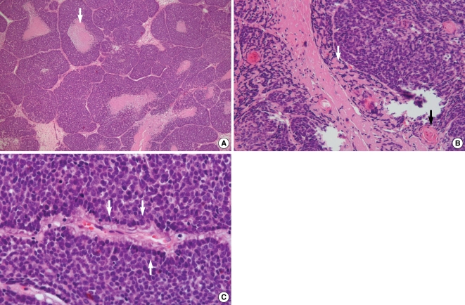
Fig. 2 Pathologic findings. (A) Irregular lobules of basaloid cells with comedo-type necrosis (arrow; H&E, ×40). (B) Abundant intercellular hyaline globules (white arrow) and multifocal keratinization (black arrow; H&E, ×200). (C) Nest of basaloid cells with peripheral palisading of the nuclei (arrows; H&E, ×400).

Fig. 3 Immunohistochemical findings showing the basaloid squamous cell features of the tumor cells (×400). (A) Nuclear immunoreactivity on the p63 staining with a brownish color (arrow). (B) Cytoplasmic and cytoplasmic membranous immunoreactivities on the high molecular weight cytokeratin staining with a brownish color (arrow). (C) No immunoreactivity on the chromogranin & CD56 staining.
Reference
-
1. Lu SY, Eng HL, Huang CC, Chien CY, Lui CC, Lin JW. Basaloid squamous cell carcinoma of the sinonasal tract: report of two cases. Otolaryngol Head Neck Surg. 2006; 5. 134(5):883–885. PMID: 16647553.
Article2. Wain SL, Kier R, Vollmer RT, Bossen EH. Basaloid-squamous carcinoma of the tongue, hypopharynx, and larynx: report of 10 cases. Hum Pathol. 1986; 11. 17(11):1158–1166. PMID: 3770734.3. Coppola D, Catalano E, Tang CK, Elfenbein IB, Harwick R, Mohr R. Basaloid squamous cell carcinoma of floor of mouth. Cancer. 1993; 10. 72(8):2299–2305. PMID: 7691390.
Article4. Raslan WF, Barnes L, Krause JR, Contis L, Killeen R, Kapadia SB. Basaloid squamous cell carcinoma of the head and neck: a clinicopathologic and flow cytometric study of 10 new cases with review of the English literature. Am J Otolaryngol. 1994; May–Jun. 15(3):204–211. PMID: 8024109.
Article5. Paulino AF, Singh B, Shah JP, Huvos AG. Basaloid squamous cell carcinoma of the head and neck. Laryngoscope. 2000; 9. 110(9):1479–1482. PMID: 10983946.
- Full Text Links
-
- Actions
-
Cited
- CITED
-
- Close
- Share
-
- Similar articles
-
- A Case of Basaloid Squamous Cell Carcinoma in the Nasal Cavity
- Basaloid-Squamous Carcinoma of the Esophagus: A case report
- Basaloid squamous cell carcinoma in the maxillary sinus
- A Case of Spontaneous Regression of Basaloid Squamous Cell Carcinoma
- A Case of Basaloid Squamous Cell Carcinoma Occurring in the Mobile Tongue
