Osteomas of the craniofacial region
- Affiliations
-
- 1Department of Oral and Maxillofacial Radiology, School of Dentistry, Pusan National University, Busan, Korea. ksnah@pusan.ac.kr
- KMID: 1449955
- DOI: http://doi.org/10.5624/isd.2011.41.3.107
Abstract
- PURPOSE
The purpose of this study was to present the clinical features of a case series of osteomas in the craniofacial region and to compare them with those described in the dental literatures.
MATERIALS AND METHODS
A retrospective study of 18 patients diagnosed with osteomas in the craniofacial region was performed. The age, gender, location, symptoms, and the radiological findings were recorded.
RESULTS
There were 13 women and 5 men from 18 years to 69 years of age (mean age, 42+/-27 years). Fourteen osteomas were found in the mandible (78%), two in frontal sinus, one in sphenoid bone, and one in maxilla.
CONCLUSION
Osteomas are benign tumors composed of mature compact bone or cancellous bone. They are essentially restricted to the craniofacial skeleton and rarely, if ever, are diagnosed in other bones.
Keyword
MeSH Terms
Figure
-
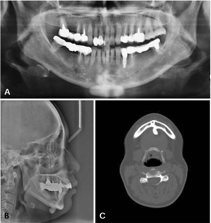
Fig. 1 Radiographic images of case I (46/F). The osteoma is located at the lingual surface of mandibular symphysis. A. Panoramic image. B. Lateral cephalometric image. C. Axial CT image.
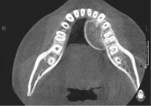
Fig. 2 Axial CT image of case II (18/F) shows the osteoma located at the lingual surface of mandibular premolar area.
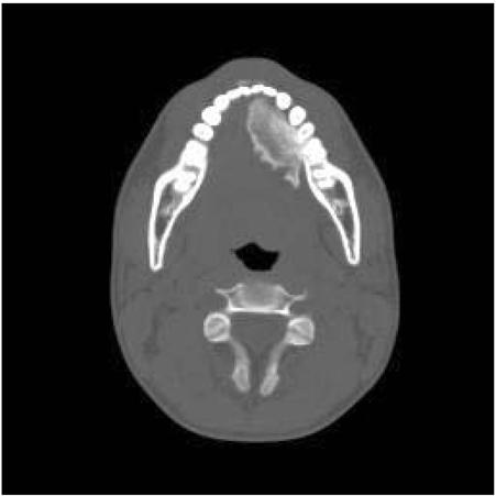
Fig. 3 Axial CT image of case III (20/M) shows the osteoma located at the lingual surface of mandibular premolar area.
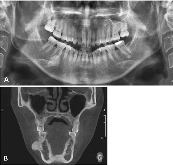
Fig. 4 A. Panoramic image of case VI (49/F) shows the osteoma located at the buccal surface of mandibular molar area. B. Coronal CT image.
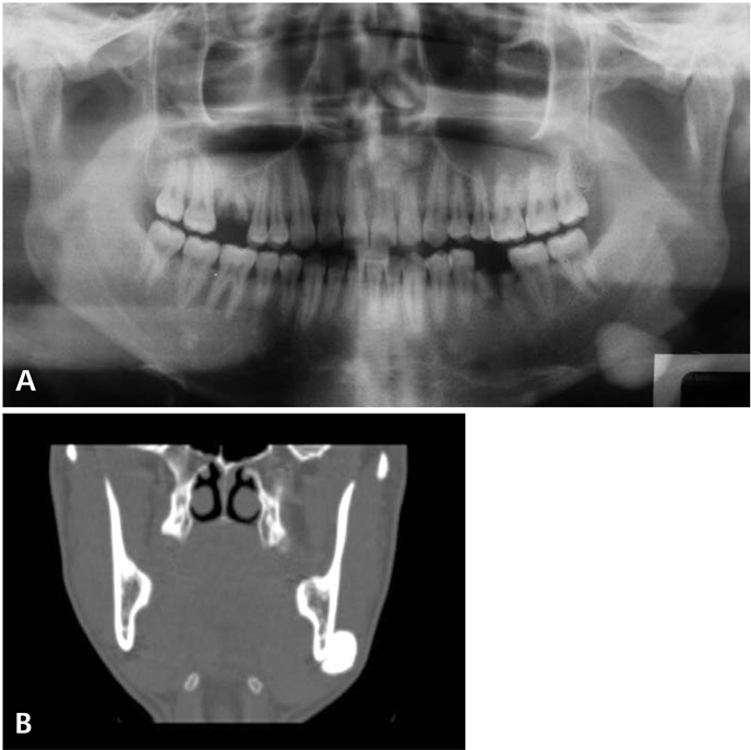
Fig. 5 A. Panoramic image of case VIII (26/M) shows the osteoma located at the buccal surface of mandibular angle area. B. Coronal CT image.
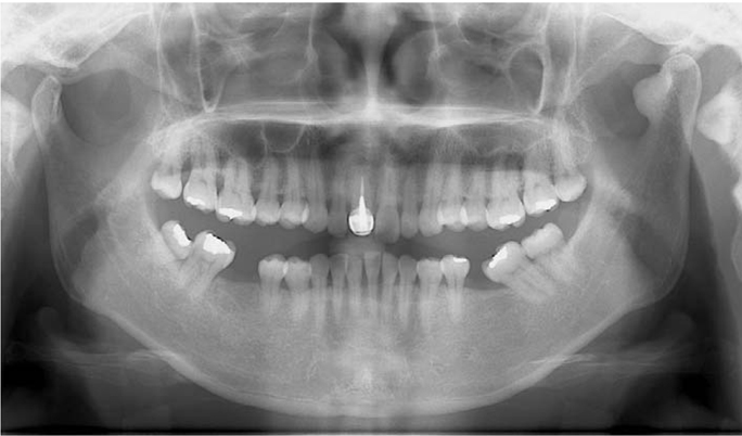
Fig. 6 Panoramic image of case XIII (44F) shows the osteoma located at the anterior surface of mandibular condyle.
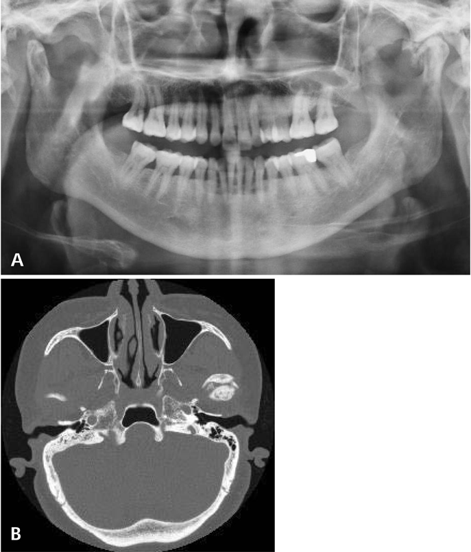
Fig. 7 A. Panoramic image of case XIV (56/F) shows the osteoma located at the anterior surface of mandibular condyle. B. Axial CT image.
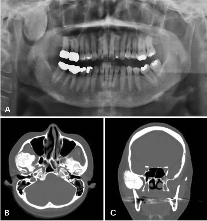
Fig. 8 A. Panoramic view of case XV (43/F) shows the osteoma located at the infra- and supra-temporal fossa of sphenoid bone. B. Axial CT image. C. Coronal CT image.
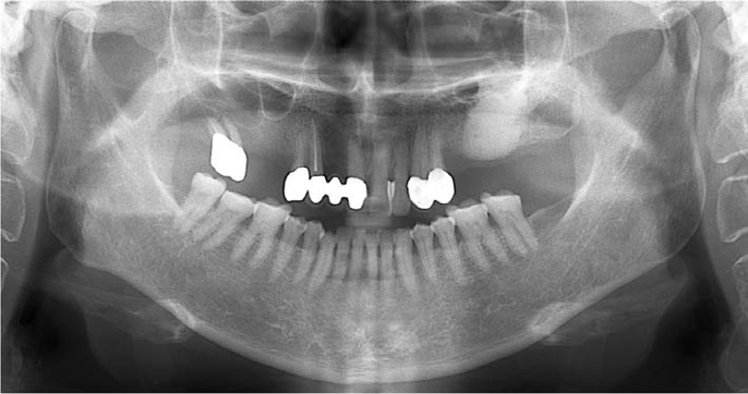
Fig. 9 Panoramic image of case XVI (69/F) shows the osteoma located at the center of left palate without any buccopalatal expansion.
Cited by 2 articles
-
Solitary peripheral osteomas of the jaws
Talita Ribeiro Tenório de França, Luiz Alcino Monteiro Gueiros, Jurema Freire Lisboa de Castro, Ivson Catunda, Jair Carneiro Leão, Danyel Elias da Cruz Perez
Imaging Sci Dent. 2012;42(2):99-103. doi: 10.5624/isd.2012.42.2.99.An unusual cause for trismus caused by mandibular coronoid osteoma: a case report
Shirin Vashishth, Kanika Garg, Prashant Patil, Venkatraman Sreenivasan
Imaging Sci Dent. 2013;43(1):45-48. doi: 10.5624/isd.2013.43.1.45.
Reference
-
1. Neville BW, Damm DD, Allen CM, Bouquot JE. Oral and maxillofacial pathology. 2002. 2nd ed. Philadelphia: WB Saunders Co;566–567.2. Piattelli A, Scarano A, Di Alberti L, Piattelli M. Osteoma of the mandible. A case report. Acta Stomatol Belg. 1995. 92:13–16.3. Larrea-Oyarbide N, Valmaseda-Castellón E, Berini-Aytés L, Gay-Escoda C. Osteomas of the craniofacial region. Review of 106 cases. J Oral Pathol Med. 2008. 37:38–42.
Article4. Kutluhan A, Salviz M, Bozdemir K, Deger HM, Culha I, Ozveren MF. Middle turbinate osteoma extending into anterior cranial fossa. Auris Nasus Larynx. 2009. 36:702–704.
Article5. Ogbureke KU, Nashed MN, Ayoub AF. Huge peripheral osteoma of the mandible: a case report and review of the literature. Pathol Res Pract. 2007. 203:185–188.
Article6. Furlaneto EC, Rocha JR, Heitz C. Osteoma of the zygomatic arch - report of a case. Int J Oral Maxillofac Surg. 2004. 33:310–311.7. Sayan NB, Uçok C, Karasu HA, Günhan O. Peripheral osteoma of the oral and maxillofacial region: a study of 35 new cases. J Oral Maxillofac Surg. 2002. 60:1299–1301.
Article8. Kashima K, Rahman OI, Sakoda S, Shiba R. Unusual peripheral osteoma of the mandible: report of 2 cases. J Oral Maxillofac Surg. 2000. 58:911–913.
Article9. Bodner L, Gatot A, Sion-Vardy N, Fliss DM. Peripheral osteoma of the mandibular ascending ramus. J Oral Maxillofac Surg. 1998. 56:1446–1449.
Article10. Ertas U, Tozoglu S. Uncommon peripheral osteoma of the mandible: report of two cases. J Contemp Dent Pract. 2003. 4:98–104.11. Longo F, Califano L, De Maria G, Ciccarelli R. Solitary osteoma of the mandibular ramus: report of a case. J Oral Maxillofac Surg. 2001. 59:698–700.
Article12. Eller R, Sillers M. Common fibro-osseous lesions of the paranasal sinuses. Otolaryngol Clin North Am. 2006. 39:585–600.
Article13. Viswanatha B. Middle turbinate osteoma. Indian J Otolaryngol Head Neck Surg. 2008. 60:266–268.
Article14. Lin CJ, Lin YS, Kang BH. Middle turbinate osteoma presenting with ipsilateral facial pain, epiphora, and nasal obstruction. Otolaryngol Head Neck Surg. 2003. 128:282–283.
Article15. Ishimaru T. Superior turbinate osteoma: A case report. Auris Nasus Larynx. 2005. 32:291–293.
Article
