Application of spherical coordinate system to facial asymmetry analysis in mandibular prognathism patients
- Affiliations
-
- 1Department of Oral and Maxillofacial Radiology, School of Dentistry, Dental Science Research Institute, Chonnam National University, Gwangju, Korea. yoonfr@chonnam.ac.kr
- 2Department of Biologic and Material Sciences, School of Dentistry, University of Michigan, Ann Arbor, USA.
- 3Department of Orthodontics, 2nd Stage of Brain Korea 21, School of Dentistry, Dental Science Research Institute, Chonnam National University, Gwangju, Korea.
- 4Department of Oral and Maxillofacial Radiology, School of Dentistry, Chonnam National University, Gwangju, Korea.
- 5Department of Orthodontics, School of Dental Medicine, Case Western Reserve University, Cleveland, USA.
- KMID: 1449953
- DOI: http://doi.org/10.5624/isd.2011.41.3.95
Abstract
- PURPOSE
The purpose of this study was to compare asymmetric mandibular prognathism individuals with symmetric mandibular prognathism individuals using a new alternate spherical coordinate system.
MATERIALS AND METHODS
This study consisted of 47 computed tomographic images of patients with mandibular prognathism. The patients were classified into symmetric and asymmetric groups. Mandibular and ramal lines were analyzed using an alternate spherical coordinate system. The length as well as midsagittal and coronal inclination angle of the lines was obtained. The bilateral differences of the spherical coordinates of the facial lines were statistically analyzed in the groups.
RESULTS
There were significant differences between the groups in bilateral difference of the length and midsagittal inclination angle of the lines (p<0.05). The bilateral difference of the length and midsagittal inclination angle of the lines has significant correlation with chin deviation (p<0.05).
CONCLUSION
The new alternate spherical coordinate system was able to effectively evaluate facial lines. The bilateral difference of lengths and midsagittal inclination of the facial lines might contribute to the facial asymmetry in mandibular prognathism individuals.
MeSH Terms
Figure
-
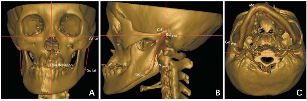
Fig. 1 A mandibular prognathism individual. Three orthogonal planes were established, and facial lines were identified. A. The chin deviation was 12.95°. The ramus lateral (RL) was identified with the most lateral points of condyle and of gonoin area. B. The longitude of the ramus (LR) was identified with the most superior point of condyle and the most inferior point of gonion area, and the ramus posterior (RP) with the most posterior points of condyle and gonion area. C. The mandibular body (MB) was identified with menton and the most posterior point of gonion area.

Fig. 2 A. An alternate spherical coordinate system (v, θ, φ) for 3D evaluation of the longitude of ramus (Cdsup-Goinf) as an example, where v is the length, θ the midsagittal inclination angle, φ the coronal inclination angle. B. This picture shows the alternate spherical coordinate system of the longitude of the ramus of the patient in Figure 1.
Cited by 4 articles
-
Normal range of facial asymmetry in spherical coordinates: a CBCT study
Suk-Ja Yoon, Rui-Feng Wang, Hee Ja Na, Juan Martin Palomo
Imaging Sci Dent. 2013;43(1):31-36. doi: 10.5624/isd.2013.43.1.31.Use of spherical coordinates to evaluate three-dimensional facial changes after orthognathic surgery
Suk-Ja Yoon, Rui-Feng Wang, Sun-Youl Ryu, Hyeon-Shik Hwang, Byung-Cheol Kang, Jae-Seo Lee, Juan M. Palomo
Imaging Sci Dent. 2014;44(1):15-20. doi: 10.5624/isd.2014.44.1.15.Deviation of landmarks in accordance with methods of establishing reference planes in three-dimensional facial CT evaluation
Kaeng Won Yoon, Suk-Ja Yoon, Byung-Cheol Kang, Young-Hee Kim, Min Suk Kook, Jae-Seo Lee, Juan Martin Palomo
Imaging Sci Dent. 2014;44(3):207-212. doi: 10.5624/isd.2014.44.3.207.A comparative study of the deviation of the menton on posteroanterior cephalograms and three-dimensional computed tomography
Hee Jin Lee, Sungeun Lee, Eun Joo Lee, In Ja Song, Byung-Cheol Kang, Jae-Seo Lee, Hoi-Jeong Lim, Suk-Ja Yoon
Imaging Sci Dent. 2016;46(1):33-38. doi: 10.5624/isd.2016.46.1.33.
Reference
-
1. Grummons DC, Kappeyne van de Coppello MA. A frontal asymmetry analysis. J Clin Orthod. 1987. 21:448–465.2. Haraguchi S, Takada K, Yasuda Y. Facial asymmetry in subjects with skeletal Class III deformity. Angle Orthod. 2002. 72:28–35.3. Ferguson JW. Cephalometric interpretation and assessment of facial asymmetry secondary to congenital torticollis. The significance of cranial base reference lines. Int J Oral Maxillofac Surg. 1993. 22:7–10.
Article4. Decker JD. Asymmetric mandibular prognathism: a 30-year retrospective case report. Am J Orthod Dentofacial Orthop. 2006. 129:436–443.
Article5. Matteson SR, Bechtold W, Phillips C, Staab EV. A method for three-dimensional image reformation for quantitative cephalometric analysis. J Oral Maxillofac Surg. 1989. 47:1053–1061.
Article6. Ono I, Ohura T, Narumi E, Kawashima K, Matsuno I, Nakamura S, et al. Three-dimensional analysis of craniofacial bones using three-dimensional computer tomography. J Craniomaxillofac Surg. 1992. 20:49–60.
Article7. Katsumata A, Fujishita M, Maeda M, Ariji Y, Ariji E, Langlais RP. 3D-CT evaluation of facial asymmetry. Oral Surg Oral Med Oral Pathol Oral Radiol Endod. 2005. 99:212–220.
Article8. Maeda M, Katsumata A, Ariji Y, Muramatsu A, Yoshida K, Goto S, et al. 3D-CT evaluation of facial asymmetry in patients with maxillofacial deformities. Oral Surg Oral Med Oral Pathol Oral Radiol Endod. 2006. 102:382–390.
Article9. Hwang HS, Hwang CH, Lee KH, Kang BC. Maxillofacial 3-dimensional image analysis for the diagnosis of facial asymmetry. Am J Orthod Dentofacial Orthop. 2006. 130:779–785.
Article10. Kwon TG, Park HS, Ryoo HM, Lee SH. A comparison of craniofacial morphology in patients with and without facial asymmetry--a three-dimensional analysis with computed tomography. Int J Oral Maxillofac Surg. 2006. 35:43–48.
Article11. Park SH, Yu HS, Kim KD, Lee KJ, Baik HS. A proposal for a new analysis of craniofacial morphology by 3-dimensional computed tomography. Am J Orthod Dentofacial Orthop. 2006. 129:600.e23–600.e34.
Article12. Baek SH, Cho IS, Chang YI, Kim MJ. Skeletodental factors affecting chin point deviation in female patients with class III malocclusion and facial asymmetry: a three-dimensional analysis using computed tomography. Oral Surg Oral Med Oral Pathol Oral Radiol Endod. 2007. 104:628–639.
Article13. You KH, Lee KJ, Lee SH, Baik HS. Three-dimensional computed tomography analysis of mandibular morphology in patients with facial asymmetry and mandibular prognathism. Am J Orthod Dentofacial Orthop. 2010. 138:540.e1–540.e8.
Article14. Thomas GB, Finney RL. Calculus and analytic geometry. 1982. 5th ed. Reading: Addison-Wesley;669–670.15. Fisher NI, Lewis T, Embleton BJJ. Statistical analysis of spherical data. 1987. Cambridge: Cambridge University Press;17–28.
- Full Text Links
-
- Actions
-
Cited
- CITED
-
- Close
- Share
-
- Similar articles
-
- Evaluation of gonial angle asymmetry after IVRO in facial asymmetry with mandibular prognathism
- Facial asymmetry with mandibular prognathism: A new trial of classification and interpretation
- Normal range of facial asymmetry in spherical coordinates: a CBCT study
- Nasal deviation in patients with mandibulo-facial asymmetry
- Osteogenesis imperfecta and combined orthodontics and orthognathic surgery: a case report on two siblings
