Lab Med Online.
2011 Apr;1(2):105-109. 10.3343/lmo.2011.1.2.7.
Evaluation of an Automated Instrument, PREVI Isola(R) for Inoculation of Body Fluids and Urine Samples onto Agar Plates
- Affiliations
-
- 1Department of Laboratory Medicine, Yonsei University College of Medicine, Seoul, Korea. deyong@yuhs.ac
- 2Research Institute of Bacterial Resistance, Yonsei University College of Medicine, Seoul, Korea.
- KMID: 1446277
- DOI: http://doi.org/10.3343/lmo.2011.1.2.7
Abstract
- BACKGROUND
In most clinical microbiology laboratories, inoculation of specimens on plates is performed manually and is a time-consuming process. The efficiency of this process can be improved by using an automated instrument. Currently, several automated instruments have been introduced for inoculation of samples. In this study, we have evaluated an automated instrument, PREVI Isola(R) (Biomerieux, France), used for inoculation of body fluids and urine specimens.
METHODS
Both manual and automated instrument methods were used to inoculate 74 body fluid and 204 urine samples. Precision was evaluated by testing 3 types of urine samples (A, 6x10(3) colony-forming units (CFU)/mL; B, 3x10(4) CFU/mL; and C, >10(6) CFU/mL) in replicates of 20. Results of the 2 methods were compared by counting the isolated colonies on agar plates after incubation. The time required for both methods was also compared.
RESULTS
The coefficient of variation (CV) of samples A, B, and C examined using the automated instrument method was 176.1%, 18.1%, and 12.6%, respectively. The sensitivity and specificity of testing body fluid samples were 77% and 100%, respectively, and those of urine samples were 87% each. The time required for testing 15 body fluid specimens and that for inoculation of each specimen was 9.7 min shorter using PREVI Isola(R) than using the manual method.
CONCLUSIONS
The results of body fluid and urine culture by inoculation using the automated instrument, PREVI Isola(R), showed relative good agreement with those obtained using the manual method. The use of PREVI Isola(R) would be expected to reduce the time and labor involved in inoculating various kinds of specimens.
MeSH Terms
Figure
-
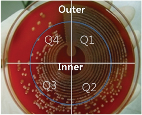
Fig. 1 The colony morphology on plate by PREVI Isola®. The agar plate is divided in 8 sections (quadrant 1-quadrant 4 and outer/inner zone).
Cited by 1 articles
-
Evaluation of Automated Specimen Inoculation for Blood Culture Samples by Use of the Previ Isola® System Compared with the Manual Method
Yeongchun Park, Jin Sang Yoon, Jimyung Kim, Gye Cheol Kwon, Sun Hoe Koo
Lab Med Online. 2016;6(1):36-40. doi: 10.3343/lmo.2016.6.1.36.
Reference
-
1. Woods GL. Automation in clinical microbiology. Am J Clin Pathol. 1992. 98:S. S22–S30.2. Bourbeau PP, Swartz BL. First evaluation of the WASP, a new auto mated microbiology plating instrument. J Clin Microbiol. 2009. 47:1101–1106.
Article3. Glasson JH, Guthrie LH, Nielsen DJ, Bethell FA. Evaluation of an automated instrument for inoculating and spreading samples onto agar pla-tes. J Clin Microbiol. 2008. 46:1281–1284.
Article4. Albers AC, Fletcher RD. Accuracy of calibrated-loop transfer. J Clin Microbiol. 1983. 18:40–42.
Article5. Chong Y, Lee K, editors. Diagnostic Microbiology. 2009. 4th ed. Seoul: Seoheung Publishing Company;58–113.6. Clinical and Laboratory Standards Institute. Evaluation of the precisionperformance of clinical chemistry devices; approved guideline. Document EP05-A2. 2004. Wayne, PA: Clinical and Laboratory Standards Institute.7. Isenberg HD, editor. Clinical microbiology procedures handbook. 2004. 2th ed. Washington, D.C.: American Society for Microbiology.8. Kass EH. Asymptomatic infections of the urinary tract. 1956. J Urol. 2002. 167:1016–1019.9. Tilton RC, Ryan RW. Evaluation of an automated agar plate streaker. J Clin Microbiol. 1978. 7:298–304.
Article10. Scarparo C, Piccoli P, Ricordi P, Scagnelli M. Evaluation of the DipStreak, a new device with an original streaking mechanism for detection, counting, and presumptive identification of urinary tract pathogens. J Clin Microbiol. 2002. 40:2169–2175.
Article
- Full Text Links
-
- Actions
-
Cited
- CITED
-
- Close
- Share
-
- Similar articles
-
- Evaluation of Automated Specimen Inoculation for Blood Culture Samples by Use of the Previ Isola(R) System Compared with the Manual Method
- Evaluation of the Impact of Automated Specimen Inoculation, Using Previ Isola, on the Quality of and Technical Time for Stool Cultures
- Erratum: Evaluation of the Impact of Automated Specimen Inoculation, Using Previ Isola, on the Quality of and Technical Time for Stool Cultures
- Evaluation of Automated Blood Culture System for Body Fluids Culture Other Than Blood
- Multicenter Evaluation of an Image Analysis Device (APAS): Comparison Between Digital Image and Traditional Plate Reading Using Urine Cultures
