Mucinous cystadenoma of the liver with ovarian-like stroma: the need for complete resection
- Affiliations
-
- 1Department of Surgery, Gospel Hospital, Kosin University College of Medicine, Busan, Korea. ymh479@kosinmed.or.kr
- 2Department of Internal Medicine, Gospel Hospital, Kosin University College of Medicine, Busan, Korea.
- KMID: 1445786
- DOI: http://doi.org/10.4174/jkss.2011.81.Suppl1.S51
Abstract
- Cystadenoma of the liver is a rare neoplasm. Although many cystadenomas are asymptomatic, symptoms can include abdominal pain, postprandial epigastric discomfort, and nausea. Dramatic changes in hepatic imaging techniques have been helpful for diagnosing cystic lesions of the liver, such as simple cyst, hydatid cyst, cystadenoma, cystadenocarcinoma, and metastatic neuroendocrine tumors. However, it remains difficult to differentiate cystadenoma from cystadenocarcinoma for multiseptated cystic hepatic lesions with papillary projection on computed tomography (CT) and magnetic resonance imaging (MRI). Here we report the case of a 47-year-old woman with several months of postprandial discomfort and abdominal fullness. CT and MRI revealed multiseptated cystic lesions with papillary excrescences. A left hemihepatectomy was performed. Histology showed a benign mucinous cystic tumor with ovarian-like stroma.
Keyword
MeSH Terms
Figure
-

Fig. 1 Contrast-enhanced computed tomography showing a cystic lesion in liver segments II and III.
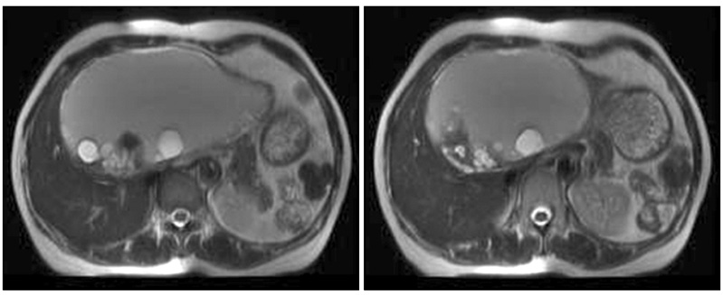
Fig. 2 Magnetic resonance imaging showing a cystic lesion in liver segments II and III with papillary projections.
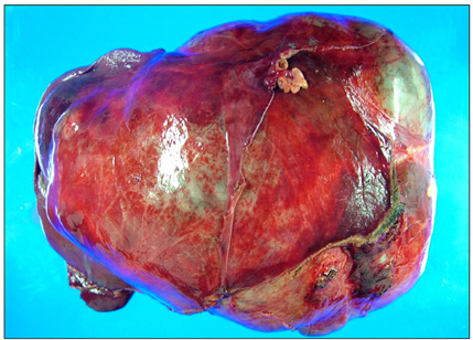
Fig. 3 Gross photograph of a typical formalin-fixed resected specimen of a biliary cystadenoma, showing a trabecular, multilocular cystic lesion with a thickened wall.
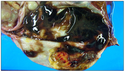
Fig. 4 Cut section of the specimen revealing a large cystic mass filled with gray-brown mucinous fluid and multiple communicating loculations of different sizes.
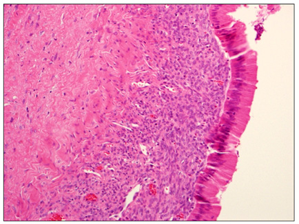
Fig. 5 The loculations were lined by columnar, cuboidal, or even flattened epithelium, which was surrounded by a layer of highly cellular, mesenchymal tissue that resembled ovarian stroma (H&E, ×400).
Reference
-
1. Thomas KT, Welch D, Trueblood A, Sulur P, Wise P, Gorden DL, et al. Effective treatment of biliary cystadenoma. Ann Surg. 2005. 241:769–773.2. Wheeler DA, Edmondson HA. Cystadenoma with mesenchymal stroma (CMS) in the liver and bile ducts. A clinicopathologic study of 17 cases, 4 with malignant change. Cancer. 1985. 56:1434–1445.3. Devaney K, Goodman ZD, Ishak KG. Hepatobiliary cystadenoma and cystadenocarcinoma. A light microscopic and immunohistochemical study of 70 patients. Am J Surg Pathol. 1994. 18:1078–1091.4. Mortelé KJ, Ros PR. Cystic focal liver lesions in the adult: differential CT and MR imaging features. Radiographics. 2001. 21:895–910.5. Suh KS, Ahn SM, Kim SW, Lee KU, Park YH, Kim ST. Biliary cystadenoma and cystadenocarcinoma. J Korean Surg Soc. 1996. 50:410–416.6. Park SJ, Lee HY, Joo SH, Kim YW, Lee SM, Hong SW. Biliary cystadenoma and cystadenocarcinoma. J Korean Surg Soc. 2007. 73:77–82.7. Sato M, Watanabe Y, Tokui K, Kohtani T, Nakata Y, Chen Y, et al. Hepatobiliary cystadenocarcinoma connected to the hepatic duct: a case report and review of the literature. Hepatogastroenterology. 2003. 50:1621–1624.8. Ishak KG, Willis GW, Cummins SD, Bullock AA. Biliary cystadenoma and cystadenocarcinoma: report of 14 cases and review of the literature. Cancer. 1977. 39:322–338.9. Regev A, Reddy KR, Berho M, Sleeman D, Levi JU, Livingstone AS, et al. Large cystic lesions of the liver in adults: a 15-year experience in a tertiary center. J Am Coll Surg. 2001. 193:36–45.10. Akwari OE, Tucker A, Seigler HF, Itani KM. Hepatobiliary cystadenoma with mesenchymal stroma. Ann Surg. 1990. 211:18–27.
