Peripancreatic Tuberculous Lymphadenopathy Mimicking Pancreatic Neoplasm: A Case Report
- Affiliations
-
- 1Department of Radiology, Sanggye Paik Hospital, Inje University College of Medicine, Korea. S2622@paik.ac.kr
- KMID: 1443507
- DOI: http://doi.org/10.3348/jksr.2011.64.6.577
Abstract
- Peripancreatic tuberculosis affecting only the lymph nodes is a rare clinical entity which usually raises serious diagnostic problems. We experienced a case of surgically proven peripancreatic tuberculous lymphadenopathy mimicking pancreas cystic neoplasm and report here on the findings of this rare condition along with a literature review.
Figure
-
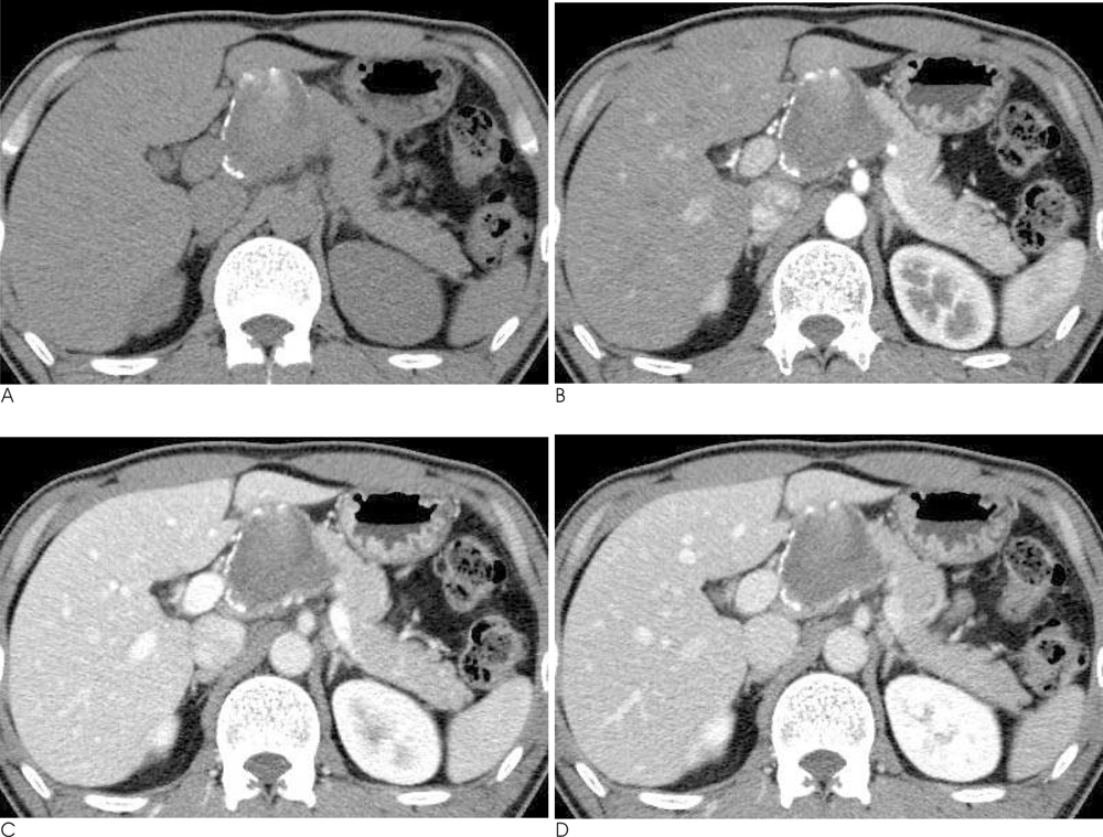
Fig. 1 Pancreatobiliary CT scan shows a 6 cm-sized, lobulating contoured mass in the pancreas head. A. Precontrast phase : a low attenuated mass with peripheral calcification and an internal highly attenuated lesion is identified. B-D. Pancreastic, portal and delayed phase: peripheral enhancement of cystic mass is noted.
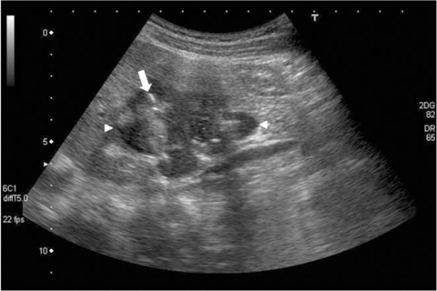
Fig. 2 Abdominal sonography shows a lobulating-contoured, heterogeneously low echoic mass (arrowhead) with internal calcification (arrow) with multiple septa in the pancreas head.
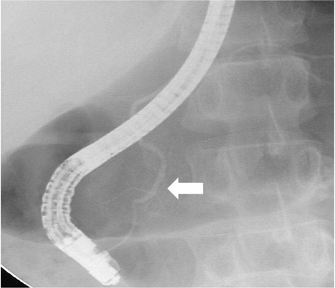
Fig. 3 ERCP shows normal main pancreatic duct (arrow).
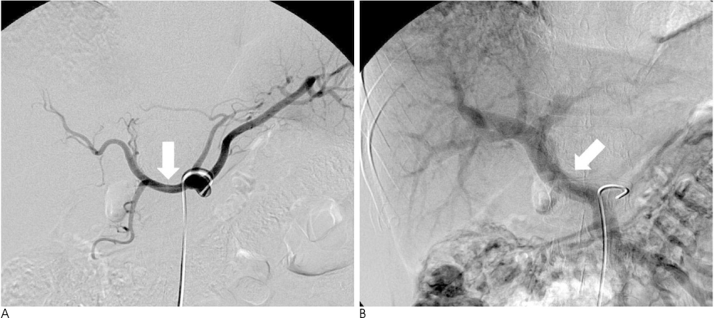
Fig. 4 Angiography shows the displacement of the common hepatic artery and proper hepatic artery (A, arrow) and main portal vein with an intact vascular wall (B, arrow).
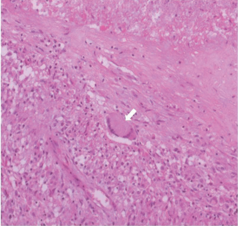
Fig. 5 Histophathogical examination revealing Langhans type giant cells (arrow) (H & E stain, ×200).
Cited by 1 articles
-
담낭염과 동반된 결핵성 담낭관 림프절염
Tae Gil Heo, Seong Woo Hong, Yeo Goo Chang, Woo Yong Lee, Haeng Jin Ohe, Kyeong Woon Choi, Yun Kyung Kang
Korean J Gastroenterol. 2021;78(4):245-248. doi: 10.4166/kjg.2021.070.
Reference
-
1. Ilhan E, Erakan N, Yildirim M, Polat AF, Çirak K, Sezgin A. Diagnostic difficulties in peripancreatic tuberculous lymphadenitis: a case report. Turk J Gastroenterol. 2005; 17:137–139.2. Sinan T, Sheikh M, Ramadan S, Sahwney S, Behbehani A. CT features in abdominal tuberculosis: 20 years experience. BMC Med Imaging. 2002; 2:3.3. Sen M, Turan M, Karadayi K, Aslan M, Elagoz S. Peripancreatic tuberculous lymphadenitis mimicking carcinoma: report of a case. Acta Chir Belg. 2004; 104:338–340.4. Kim SY, Kim MJ, Chung JJ, Lee JT, Yoo HS. Abdominal tuberculous lymphadenopathy: MR imaging findings. Abdom Imaging. 2000; 25:627–632.5. Pereira JM, Madureira AJ, Vieira A, Ramos I. Abdominal tuberculosis: imaging features. Eur J Radiol. 2005; 55:173–180.6. Akhan O, Pringot J. Imaging of abdominal tuberculosis. Eur Radiol. 2002; 12:312–323.7. Turan M, Sen M, Koyuncu A, Aydin C, Elaldi N, Arici S. Pancreatic pseudotumor due to peripancreatic tuberculous lymphadenitis. Pancreatology. 2002; 2:561–564.8. Weiss ES, Klein WM, Yeo CJ. Peripancreatic tuberculosis mimicking pancreatic neoplasia. J Gastrointest Surg. 2005; 9:254–262.9. Cherian JV, Somasundaram A, Ponnusamy RP, Venkataraman J. Peripancreatic tuberculous lymphadenopathy. An impostor posing diagnostic difficulty. JOP. 2007; 8:326–332.10. Aston NO. Abdominal tuberculosis. World J Surg. 1997; 21:492–499.
- Full Text Links
-
- Actions
-
Cited
- CITED
-
- Close
- Share
-
- Similar articles
-
- Advantages of incremental dynamic CT in the evaluation of pancreatic and peripancreatic lesions
- A Case of an Infected Aneurysm in the Thoracic Aorta Mimicking Tuberculous Lymphadenopathy
- Computed Tomographic Differential Diagnosis of Cervical Lymphadenopathy: Tuberculous versus Metastatic
- Solitary Intra-abdominal Tuberculous Lymphadenopathy Mimicking Duodenal GIST
- Peripancreatic Lesions that Need to be Differentiated from Pancreatic Tumors
