A Case of Pulmonary Capillary Hemangiomatosis-Like Lesion Followed for Seven Years
- Affiliations
-
- 1Division of Pulmonary and Critical Care Medicine, Department of Internal Medicine, The Catholic University of Korea College of Medicine, Seoul, Korea.
- 2Division of Pulmonary and Critical Care Medicine, Department of Internal Medicine, Armed Forces Capital Hospital, Seongnam, Korea.
- 3Department of Radiology, Chonnam National University Hospital, Gwangju, Korea.
- 4Division of Pulmonary and Critical Care Medicine, Department of Internal Medicine, Wonkwang University Sanbon Hospital, Wonkwang University College of Medicine, Gunpo, Korea. hikim61@hotmail.com
- KMID: 1442854
- DOI: http://doi.org/10.4046/trd.2011.70.3.242
Abstract
- Pulmonary capillary hemangiomatosis (PCH) is a rare disease of unknown etiology that is characterized by nodules composed of infiltrating capillary blood vessels. Herein, we describe a case of a PCH-like lesion that was detected by chest computed tomography. Transthoracic needle aspiration resulted in life-threatening hemorrhage. The patient was followed for seven years. He remained in good health and a follow up image showed little interval change.
MeSH Terms
Figure
-
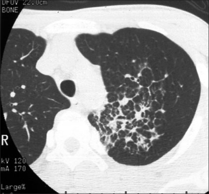
Figure 1 HRCT showed ill-defined nodular densities and interlobular and intralobular septal thickenings in LUL. HRCT: high resolution computed tomography.
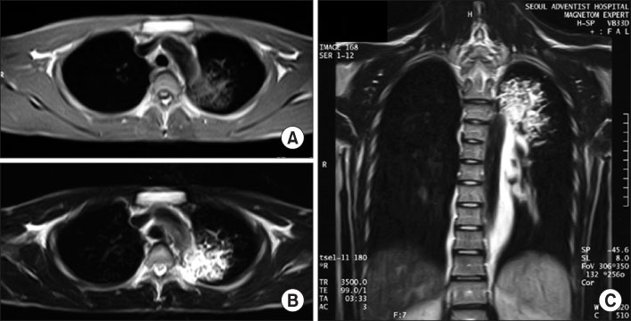
Figure 2 MRI showed reticular shaped iso-signal intensity of T1WI (A) and mottled reticular shaped high signal intensity on T2WI (B). Dysplasia of branching vessel originating from thoracic aorta was also noted on T2WI (C). MRI: magnetic resonance image; WI: weighted image.

Figure 3 CT-guided aspiration was performed in LUL. CT: computer tomography; LUL: left upper lobe.
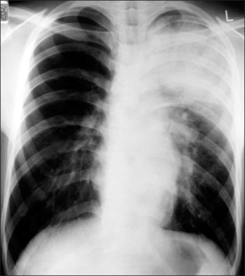
Figure 4 Chest X-ray after TTNA showed newly developed hazy density in LUL. TTNA: transthoracic needle aspiration; LUL: left upper lobe.
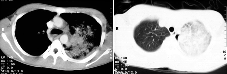
Figure 5 CT showed ill-defined air space consolidation in LUL. CT: computer tomography; LUL: left upper lobe.

Figure 6 Chest X-ray showed improvement of consolidation in LUL. LUL: left upper lobe.
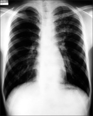
Figure 7 Chest X-ray showed further improvement of consolidation in LUL. LUL: left upper lobe.
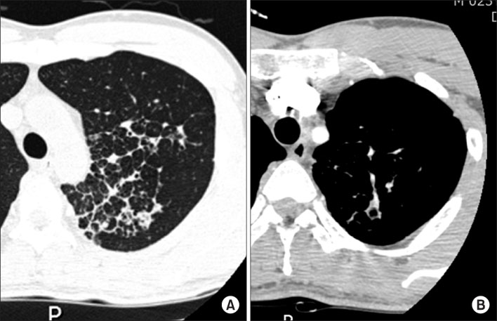
Figure 8 CT showed interlobular septal thickening (A) with enhanced vascular structure (B) in LUL. CT: computer tomography; LUL: left upper lobe.
Reference
-
1. Tron V, Magee F, Wright JL, Colby T, Churg A. Pulmonary capillary hemangiomatosis. Hum Pathol. 1986. 17:1144–1150.2. Fernández-Alonso J, Zulueta T, Reyes-Ramirez JR, Castillo-Palma MJ, Sanchez-Román J. Pulmonary capillary hemangiomatosis as cause of pulmonary hypertension in a young woman with systemic lupus erythematosus. J Rheumatol. 1999. 26:231–233.3. Case records of the Massachusetts General Hospital Weekly clinicopathological exercises. Case 38-2000. A 45-year-old woman with exertional dyspnea, hemoptysis, and pulmonary nodulas. N Engl J Med. 2000. 343:1788–1796.4. Lippert JL, White CS, Cameron EW, Sun CC, Liang X, Rubin LJ. Pulmonary capillary hemangiomatosis: radiographic appearance. J Thorac Imaging. 1998. 13:49–51.5. de Perrot M, Waddell TK, Chamberlain D, Hutcheon M, Keshavjee S. De novo pulmonary capillary hemangiomatosis occurring rapidly after bilateral lung transplantation. J Heart Lung Transplant. 2003. 22:698–700.6. Dufour B, Maître S, Humbert M, Capron F, Simonneau G, Musset D. High-resolution CT of the chest in four patients with pulmonary capillary hemangiomatosis or pulmonary venoocclusive disease. AJR Am J Roentgenol. 1998. 171:1321–1324.7. Hansell DM. Small-vessel diseases of the lung: CT-pathologic correlates. Radiology. 2002. 225:639–653.8. El-Gabaly M, Farver CF, Budev MA, Mohammed TL. Pulmonary capillary hemangiomatosis imaging findings and literature update. J Comput Assist Tomogr. 2007. 31:608–610.9. Kothari SS, Jagia P, Gupta A, Singh N, Ray R. Images in cardiovascular medicine. Pulmonary capillary hemangiomatosis. Circulation. 2009. 120:352–354.10. Lawler LP, Askin FB. Pulmonary capillary hemangiomatosis: multidetector row CT findings and clinico-pathologic correlation. J Thorac Imaging. 2005. 20:61–63.11. Eltorky MA, Headley AS, Winer-Muram H, Garrett HE Jr, Griffin JP. Pulmonary capillary hemangiomatosis: a clinicopathologic review. Ann Thorac Surg. 1994. 57:772–776.12. Ishii H, Iwabuchi K, Kameya T, Koshino H. Pulmonary capillary haemangiomatosis. Histopathology. 1996. 29:275–278.13. Almagro P, Julià J, Sanjaume M, González G, Casalots J, Heredia JL, et al. Pulmonary capillary hemangiomatosis associated with primary pulmonary hypertension: report of 2 new cases and review of 35 cases from the literature. Medicine (Baltimore). 2002. 81:417–424.14. al-Fawaz IM, al Mobaireek KF, al-Suhaibani M, Ashour M. Pulmonary capillary hemangiomatosis: a case report and review of the literature. Pediatr Pulmonol. 1995. 19:243–248.15. Takiguchi Y, Uruma T, Hiroshima K, Motoori K, Watanabe R, Hamaoka T, et al. Stable pulmonary capillary haemangiomatosis without symptomatic pulmonary hypertension. Thorax. 2001. 56:815–817.
- Full Text Links
-
- Actions
-
Cited
- CITED
-
- Close
- Share
-
- Similar articles
-
- A Case of pulmonary cavernous hemangiomatosis presented with right shoulder pain
- Diffuse Hemangiomatosis in the Intra-Abdominal Cavity Mimicking Peritoneal Metastasis: A Case Report
- A Case of Benign Neonatal Eruptive Hemangiomatosis
- Diffuse Neonatal Hemangiomatosis with Association of Massive Osteolysis and Arteriovenous Fistulae: An autopsy case
- Diffuse Neonatal Hemangiomatosis with Association of Massive Osteolysis and Arteriovenous Fistulae: An autopsy case
